Formulated diagnosis
ICD-10: C16.9 (malignant neoplasm of stomach, unspecified); ICD-O morphology 8936/3, gastrointestinal stromal tumour.
Diagnosis: Gastrointestinal stromal tumour (GIST) of the stomach, KIT exon 11 deletion (Q556_V559>H), with metastases to the liver (bilobar, dominant right-lobe mass) and peritoneum, stage IV. Initially (08/2022) diagnosed and treated as leiomyosarcoma of the uterine body T2NxM1; diagnosis revised 04–06/2023 on histology (DOG-1, CD117 positive; Israeli pathology revision of all samples). Concomitant: uterine leiomyomas; right ovarian endometrioma; type 2 diabetes; iron-deficiency anaemia.
08/2022 — liver-metastasis biopsy read as metastatic uterine leiomyosarcoma; bilobar liver masses, paragastric mass 51 × 46 mm, pelvic masses.
09/2022 – 01/2023 — status after 4 cycles gemcitabine + docetaxel + bevacizumab: progression in the liver (MRI 10/2022, CT 12/2022). Oncomine (12/2022): KIT exon 11 deletion (VAF 42.5 %), subclonal KIF5B–MET fusion, TMB low, MSS; germline panel negative.
05–12/01/2023 — pazopanib 800 mg, stopped on day 8: posterior reversible encephalopathy syndrome with left occipital haemorrhage, hospitalised 13–25/01/2023, resolved; CT 09/02/2023 with 12 % regression.
01/2023 — repeat liver biopsy: reported as metastasis of uterine leiomyosarcoma.
02–03/2023 — status after 3 cycles trabectedin: progression on PET-CT 11/04/2023.
04–06/2023 — EUS-FNA of the paragastric mass and liver core biopsy: GIST of the stomach, GIST metastasis in the liver (CSD, 21–26/04/2023); Israeli revision 04/06/2023 confirms GIST in all samples.
03/05/2023 — imatinib 400 mg/day started. 08/2023 — marked metabolic response (PET-CT). 03/2024 — stable. 05/2025 — stable, "PET negative", cystic residues.
06–09/2026 — PET-CT 19/06/2026: return of enhancing, FDG-active mural components in the known liver lesions after 3 years of imatinib (started 03.05.2023) (LISOD re-read 08/07/2026; Israeli expert reading 08/09/2026: metabolic progression, left-lobe lesion 42 × 36 mm, right-lobe mass ~15 × 10 cm, no bone lesions).
08/2026 — FoundationOne Liquid CDx (ctDNA fraction 32 %): KIT exon 11 Q556_V559>H (17.0 %) plus secondary KIT exon 13 V654A (8.8 %) and exon 18 A829P (4.0 %) — polyclonal imatinib resistance.
08/09/2026 — recommendation: sunitinib 50 mg 4/2 (regorafenib 160 mg d1–21 as an option); not yet started; imatinib continues. 10/09/2026 — thermal ablation of the left-lobe lesion judged contraindicated by Dr. O. Korshak (interventional radiology, Shalimov Centre, Kyiv): contact with the left portal branch and bile duct; other local options under discussion. 17/09/2026 — Dr. E. Tahover (GIST oncologist, Tel Aviv Sourasky): imatinib has failed, start a second-line TKI now (sunitinib or ripretinib; regorafenib an alternative); liver-directed interventional treatment first. 18/09/2026 — Dr. D. Fedorov (liver surgeon): two-stage liver resection feasible and potentially radical; fresh PET-CT, then workup.
23/09/2026 — PET-CT (KMKOC): further metabolic progression in multiple liver foci versus 19/06/2026, SUVmax up to 4.9; left-lobe active area confluent, up to 35 × 54 mm; active focus up to 15 × 24 mm in segment S8; gastric and uterine lesions stable; oesophageal uptake SUVmax 2.0–3.2 (endoscopy advised); no bone lesions.
30/09/2026 — regorafenib (Stivarga 40 mg tablets × 84 = one 21-day cycle at 160 mg/day) dispensed at LISOD; not yet started. Baseline blood tests the same day (results).
Current situation (as of 30 September 2026)
Status as of 20 October 2026 (latest first)
- 20.10.2026 — therapy (family report, 20.10.2026): imatinib 400 mg/day since 03.05.2023: stopped 20.10.2026 before the operation; restart planned a few days after the operation (timing to be discussed); regorafenib dispensed 30.09.2026, not started, not allowed currently.
- OPINION 20.10.2026 — Dr D. Fedorov (opinion, telephone): the planned operation is a right hemihepatectomy (major liver resection); the stomach tumour is removed in the same operation where possible, unless it would need an extra-large gastric resection — then it is left for the second stage; hospital stay at least 1–1.5 weeks; risks named: deviation from the plan, rupture of the tumour during the operation (thin walls, necrosis inside), bleeding, wound infection. The patient was already in hospital (family statement). No operation date was stated in this call (summary).
- OPINION 16.10.2026 — Dr D. Fedorov (opinion, telephone; reporting the decision of the Shalimov Centre tumour board, MOK): the MOK approved surgery; first stage most likely a right hemihepatectomy (the right-lobe lesion has a fluid component that could rupture; most of the right lobe is non-functioning tumour); the stomach tumour removed in the same operation if a local resection is enough (decided at intra-operative revision); second stage: the left-lobe lesion; no local treatment of the left-lobe lesion now. On the ascitic cytology he considers the earlier category V finding over-diagnosis against the background of the cancer diagnosis (presumably the first report). Operation possible "this Wednesday" if the patient agrees (INTERPRETATION 21.10.2026, computed from the call date); the written MOK conclusion was due on Monday 19.10.2026 (not on file); the team's talk with the patient will be the signing of the consent; imaging about two weeks after surgery to assess liver regeneration (summary).
- Imatinib before the operation — resolved (family report, 20.10.2026): imatinib stopped on 20.10.2026 before surgery; restart a few days after surgery — timing to be discussed with the surgeon/oncologist. Earlier: family report of 14.10.2026 — taken and continued until the operation; OPINION Dr D. Fedorov, 16.10.2026, asked whether she will take imatinib: "not now"; after the operation she returns to it once her condition allows (summary) — consistent with the stop before surgery.
- FACT 14.10.2026 — Ascitic fluid cytology: category V (Shalimov, 06.10) not reproduced on review of the same slides (CSD LAB, 14.10: AUS) — malignancy not established[196][197]; see Ascitic fluid cytology.
- FACT 13.10.2026 — discharge summary, Shalimov Centre (stay 05.10–13.10.2026; medical card 9577/2026): diagnosis as written "C78.7 GIST of the stomach cT2N0M1 (hep), KIT exon 11 mutation. Stage IV. Morphologically confirmed 13.10.2026"; comorbid: uterine leiomyoma, endometrioma of the right ovary, type 2 diabetes, iron-deficiency anaemia. EGD 02.10.2026: erythematous gastropathy, duodeno-gastric bile reflux (the oesophagus is not described). Family report (20.10.2026): she had a cold with a mild sore throat in the days of the 23.09.2026 PET-CT, whose report states that the oesophageal uptake may be of inflammatory origin. Colonoscopy 02.10.2026: single (3) neoplasias of the colon (histology not stated). Thermal ablation of the left-lobe focus "judged technically inadvisable". Post-operative period without complications; recommended review at the Centre on the results of the oncology consilium, to decide on radical surgery. The summary contains no ascites, peritoneal or cytology information (English translation[201] ).
- OPINION 13.10.2026 — Dr D. Fedorov (opinion, Telegram): repeat the cytology; if it is confirmed, surgery is not indicated. The review did not confirm it; his comment on that is not documented (messages).
- FACT 05.10.2026 — diagnostic laparoscopy, Shalimov Centre (pathology report 13.10.2026): ultrasound-guided trepan biopsy of the liver lesion in segments II–III — GIST metastasis, spindle-cell, low grade, CD117+, DOG-1+, Ki-67 2 %; incisional biopsy of the gastric lesion — hyalinised tissue without viable tumour cells, which may be a consequence of therapy (translation[195]; details).
- INTERPRETATION The biopsied liver lesion is the left-lobe lesion L1 by location only; the report gives no size or image.
- FACT 30.09.2026 — therapy: imatinib 400 mg/day since 03.05.2023, taken currently and continued until the operation (resumed 06.10.2026, the day after the laparoscopy; family report, 14.10.2026); regorafenib dispensed 30.09.2026, not started — held because surgery is expected soon (chosen at LISOD; family report, 14.10.2026). OPINION On 29.09.2026 Prof. A. Czarnecka, Onkolmed, opinion by phone: "not a bad choice" (summary).
- FACT 23.09.2026 — PET-CT (KMKOC): FDG activity increased in multiple liver foci versus 19.06.2026, SUVmax up to 4.9[187]. INTERPRETATION Volumes from this site's three-date model (3D viewer): liver 2.74 L; dominant right-lobe mass R1 about 1.2 L, stable (1.21 → 1.26 → 1.20 L, May 2025 → Jun 2026 → Sep 2026); S8 dome lesion 79 mL (68 → 71 → 79 mL) with a new FDG-active focus; left-lobe lesion L1 54 mL (21 → 33 → 54 mL), FDG-active and confluent; gastric formation GF FDG-cold, 37 mL on the non-contrast September CT (scale less certain; 30 and 27 mL before). Correlation: the FDG-active L1 is viable GIST on biopsy; the FDG-cold, calcified GF had no viable tumour in the sample. Ascites was not described on this scan.
- FACT 20.08.2026 — resistance: KIT exon 13 V654A and exon 18 A829P in blood (FoundationOne Liquid CDx)[75].
- FACT Background (2022): spread recorded in 2022, verbatim: «T2NxM1 (печінка, саркоматоз черевної порожнини, великого чепця)» — "liver, sarcomatosis of the abdominal cavity, greater omentum" (anamnesis line of the MRI report of 14.10.2022, National Cancer Institute of Ukraine[5]; Original · UA English translation). No 2022 document on file uses the words "peritoneal metastases".
Pending: cytology of new ascitic fluid with a cell block and IHC (CD117/DOG-1), if fluid is available (the second laboratory read of the existing slides, which Dr Fedorov asked for, is done: CSD LAB, 14.10.2026; whether a new puncture is feasible is for the treating team); laparoscopy operation protocol (Shalimov Centre); oncology consilium (treating oncologists and surgeons) — OPINION held: surgery approved (Dr Fedorov, 16.10.2026); its written conclusion (due 19.10.2026) is not on file; imatinib: stopped 20.10.2026 before surgery; restart a few days after surgery — timing to be discussed with the surgeon/oncologist (family report, 20.10.2026); regorafenib: not started, held because surgery is expected soon (family report, 14.10.2026); not allowed currently (family report, 20.10.2026); timing for the treating oncologist; endoscopy for the oesophageal uptake advised on 23.09.2026 (status not documented) — EGD done on 02.10.2026 per the discharge summary of 13.10.2026[201] (erythematous gastropathy, duodeno-gastric bile reflux; the oesophagus is not described in the summary); family report 20.10.2026: she had a cold with a mild sore throat in those days; open for the treating team; nature of the colon neoplasias seen at colonoscopy on 02.10.2026 ("single (3)"; histology not stated in the summary — to request); genetics only on operative material, per the oncologist Dr O.M. Latsinnikova (opinion relayed in chat, 13.10.2026); whether a third Shalimov cytology slide exists; trial eligibility and tissue biobanking under consideration (living-tissue banking in Israel not pursued: family report, 20.10.2026, summary).
Extent of the current progression. This is not a single small nodule, and it grew between June and September:
- FACT PET-CT 23.09.2026 (KMKOC): FDG activity increased in multiple liver foci compared with 19.06.2026, SUVmax up to 4.9 — progression of the underlying disease.
- FACT Left lobe (segments II/III): the active area is now confluent, up to 35 × 54 mm (June CT lesion 46 × 48 × 42 mm, 27 mL, expert reading 42 × 36 mm). On the June contrast CT it touches the left portal branch and the left hepatic duct.
- FACT Right lobe: an active focus up to 15 × 24 mm in segment S8, inside the ametabolic area of the dominant mass (now 98 × 148 mm). In June the right-lobe activity was a plaque on the wall of that mass.
- INTERPRETATION Own volumetry (analysis): FDG-active tissue in the left lobe 19 → 36 mL (2.2 → 3.4 × liver background); new 4 mL focus in S8; active wall of the right-lobe mass 6 → 10 mL.
- FACT ctDNA tumour fraction 32 % (FoundationOne Liquid CDx, 20.08.2026) INTERPRETATION is high and points to a substantial amount of active tumour, carrying two resistance clones (KIT exon 13 V654A and exon 18 A829P).
Patient. Anastasia Zherlitsyna, born 25.04.1976 (50), Kyiv, Ukraine.
Diagnosis. Metastatic gastric GIST with bilobar liver metastases; treated as uterine leiomyosarcoma T2NxM1 from 28.09.2022 until the April–May 2023 biopsies established GIST (DOG-1 and CD117 positive; Israeli revision 04.06.2023 confirmed GIST in all three samples). Uterine leiomyomas and a right ovarian endometrioma are benign bystanders.
Molecular status (most important). Driver KIT exon 11 deletion Q556_V559>H (Oncomine, Dec 2022, VAF 42.5 %). FoundationOne Liquid CDx of 20.08.2026, ctDNA tumour fraction 32 %: exon 11 driver at VAF 17.0 % plus two new KIT resistance mutations — exon 13 V654A (8.8 %) and exon 18 A829P (4.0 %), in different kinase domains: polyclonal imatinib resistance. The report flags imatinib, sunitinib and avapritinib as resistant and lists regorafenib as NCCN category 1. Which drug covers which clone: drug table; every variant from both tests: genomic findings.
Sites of disease (PET-CT 23.09.2026, KMKOC). Multiple ametabolic liver lesions with metabolic activity growing along the contour and inside some of them, SUVmax up to 4.9; dominant right-lobe mass 98 × 148 mm (June ~144 × 104 mm); left-lobe active area up to 35 × 54 mm, confluent; active focus up to 15 × 24 mm in segment S8; calcified FDG-negative gastric lesion 43 × 32 mm, unchanged; uterine lesions stable; new mild–moderate oesophageal uptake (SUVmax 2.0–3.2, possibly inflammatory — endoscopy advised); no bone lesions. June 2026 (contrast CT, expert reading 08.09.2026): left-lobe lesion 42 × 36 mm (own measurement 46 × 48 × 42 mm, 27 mL), in contact with the left portal branch.
Treatment. Imatinib 400 mg/day since 03.05.2023, continuing; taken currently and continued until the operation (resumed 06.10.2026, the day after the laparoscopy; family report, 14.10.2026). Stopped on 20.10.2026 before the operation; restart planned a few days after the operation, timing to be discussed (family report, 20.10.2026). On 30.07.2026 and 08.09.2026 the LISOD oncologist recommended sunitinib 50 mg, 28 days on / 14 off (regorafenib 160 mg days 1–21 q28 as an alternative) — sunitinib was not started. Regorafenib (Stivarga 40 mg tablets × 84, one 21-day cycle at 160 mg/day) was dispensed at LISOD on 30.09.2026 and has not been started yet (as of 30.09.2026); not started; held because surgery is expected soon (family report, 14.10.2026); not allowed currently (family report, 20.10.2026); baseline blood tests the same day (results). On 17.09.2026 Dr. Tahover (Ichilov) urged starting a second-line TKI without further delay.
Safety context. PRES on pazopanib in January 2023 with left occipital haemorrhage and multifocal vasoconstriction, largely resolved by June 2023 — potent VEGFR inhibitors carry extra risk. Baseline echocardiogram 30.07.2026 normal (EF 67 %). Comorbidities on record: type 2 diabetes, iron-deficiency anaemia.
Open clinical question. Local treatment of the FDG-active left-lobe lesion: on 10.09.2026 Dr. Oleksandr Korshak (interventional radiology, Shalimov Centre of Surgery and Transplantology, Kyiv) judged thermal ablation (RFA/MWA) contraindicated because the lesion contacts the left portal branch and the accompanying left bile duct (imaging analysis); TARE, TACE/DEB-TACE, SBRT, IRE, resection of segments II–III and non-thermal histotripsy (HistoSonics, newly CE-approved; opinion to be requested from the manufacturer, as Dr. Korshak suggested) are under discussion, with imatinib continued — an approach NCCN and ESMO allow to be considered for limited progression on imatinib. The PET-CT of 23.09.2026 adds an active focus in segment S8 of the right lobe, which any local plan must cover as well; S8 lies within the part removed by a right hemihepatectomy, the first stage of the two-stage resection Dr. Fedorov proposed on 18.09.2026 (guideline basis, ablation opinion).
Which 3D model to use
This page has one 3D model to use and three older ones.
START HEREThree-date liver model — open. It shows the liver, the tumours and their PET-active (metabolically active) parts on 13.05.2025, 19.06.2026 and 23.09.2026. Switch dates with the buttons. Press Compare to see the earlier scan as an outline on the current one. Hover any part to see what the doctors' reports say about it. It is an AI-assisted reconstruction, not a radiology report. Less certain on 23.09.2026 (no contrast): liver segments, veins and the portal vein are estimated; the stomach and the gastric tumour were outlined by one reader only. It adds each tumour separately (R1, L1, S8, P7, S1), the stomach tumour, the veins, the active tissue and a comparison between dates.
Older 3D models (for reference):
- Liver model of 19.06.2026 and liver model of 23.09.2026: one scan each; older version; shows fewer structures; colours differ (green = right-lobe mass, cyan = left-lobe lesion, yellow = active tissue); do not use for measurements. The dotted orange line is a guessed path of the bile duct, drawn from the June contrast CT; it is not measured. The red ball is where the left lesion touches a portal-vein branch on the June scan; the September scan (no contrast) cannot show this.
- Whole-body model of 23.09.2026: lesions in colour, organs and bones faint; for the overview of all sites.
- Gastric and pelvic masses over time: the gastric-wall tumour on eleven scans and the uterus on six PET-CTs, side by side at one scale.
Liver 3D across three CTs: 13.05.2025 / 19.06.2026 / 23.09.2026
Whole-body model of all lesions (PET-CT 23.09.2026)
- Operate now? Does the unresolved cytology (category V; AUS on review) stop surgery, or is it settled by peritoneal inspection and frozen-section washings at the start of surgery?
- Order and staging. Right hemihepatectomy (R1 with the S8 lesion) first or L1 first; one stage or two; and how is R1 — the 1.2 L right-lobe mass with FDG-active wall plaques — handled: resection only, or is drainage/aspiration an option?
- L1 technique and operator. For L1 — 54 mL on the 23.09.2026 consensus model (4.9 × 3.9 × 5.9 cm), biopsy-proven viable GIST (05.10.2026) [195], 0–1 mm from the left portal branch and the left bile duct — which option is feasible here, where and by whom: resection of the left lateral section (segments II–III), irreversible electroporation (NanoKnife), histotripsy (HistoSonics), SBRT, or transarterial treatment (DEB-TACE/TARE) of the segment II/III feeding artery?
Background
As first put (before the biopsies of 05.10.2026; the 15 × 24 mm S8 focus of the report is 14 × 23 mm on the consensus model): Physical removal of the new focus, with imatinib continued. Thermal ablation has been declined because the segment II/III lesion (June 42 × 36 mm; FDG-active area 35 × 54 mm, confluent, on 23.09.2026) abuts the left portal branch and the left hepatic duct; the 23.09.2026 PET-CT adds an active 15 × 24 mm focus in segment S8. Which option does the board recommend and where can it be done: resection of the left lateral section (segments II–III), irreversible electroporation (NanoKnife), histotripsy (HistoSonics, newly approved in Europe; HCA price list 11.09.2026), SBRT, or transarterial treatment (DEB-TACE/TARE) of the segment II/III feeding artery — and how should the active rim of the right-lobe mass be handled?
- Same-session targets. Are the S8 dome lesion (79 mL) with its new 14 × 23 mm active focus, the R1 wall plaques and the calcified gastric primary [195] addressed in the same operation, or left or treated separately?
- Operator, venue, window. Who operates, where, and in which window (from 19.10.2026? OPINION), and is the contrast CT with volumetry done before the first operation (last contrast study 19.06.2026 [70])?
- Imatinib and regorafenib. Imatinib stopped 20.10.2026 before surgery: confirm the restart date after the operation; regorafenib not allowed now: timing of regorafenib or an alternative after the local step?
- PRES safety. After haemorrhagic PRES (2023) [22]: peri-operative blood-pressure target, anaesthesia plan, and which VEGFR-active drugs (sunitinib, regorafenib) to avoid around surgery and SBRT?
- Response check. Which check after each step — contrast CT at 4–6 weeks, PET-CT at 3 months, repeat ctDNA — and who orders and reads it?
- Next systemic line or trial. Which drug or trial would cover both resistance clones (KIT exon 13 V654A and exon 18 A829P) at the lowest neurovascular risk after her PRES — sunitinib, regorafenib, ripretinib, velzatinib (StrateGIST 3), bezuclastinib + sunitinib or pimitespib — and at what point: now, or only after local control fails?
Background
As first put (27.09.2026): New systemic drug or trial, if and when needed. Both resistance clones (KIT exon 13 V654A and exon 18 A829P) must be covered; each later TKI line gives a shorter time to the next progression with more toxicity; she had PRES with intracerebral haemorrhage on a VEGFR inhibitor. Full comparison in the drug table.
Option Covers V654A / A829P Expected benefit Main risk for her Access / cost Sunitinib 50 mg 4/2 (prescribed) yes / no median PFS 5–15 months by clone hypertension, VEGFR class — PRES history prescribed 30.07 and 08.09.2026, not started — regorafenib chosen instead Regorafenib 160 mg d1–21 yes (VOYAGER ctDNA) / yes median PFS about 5 months hypertension, hand–foot NCCN category 1; chosen — dispensed 30.09.2026, not yet started Ripretinib 150 mg weak / strong 4–6 months; equal to sunitinib in the co-mutated subgroup lowest neurovascular risk of the TKIs about USD 36,600 per month (Israeli price ₪110,986 per 90 tablets, 09.2026) Velzatinib (IDRX-42), StrateGIST-1 yes / yes 2nd line 13.7 months, 3rd line 9.2 months selective KIT, low VEGFR trial: StrateGIST 3 (velzatinib vs sunitinib after imatinib) recruiting as of 27.09.2026, incl. Warsaw (Dr P. Rutkowski), Poznań, Kielce, Essen, Berlin, Mannheim, Budapest; randomised 1:1, so a 50 % chance of sunitinib Bezuclastinib + sunitinib (Peak) yes / yes phase 3 (ASCO 2026, n = 413): PFS 16.5 vs 9.2 months vs sunitinib alone, HR 0.50; ORR 46 vs 26 % sunitinib risk retained FDA decision due 30.11.2026; Peak no longer enrolling in Europe Pimitespib (HSP90 inhibitor) mutation-agnostic PFS 2.8 vs 1.4 months, stabilisation only diarrhoea, night blindness; no VEGFR activity Japan only; import about €17,700 per cycle Question: which of these would the board choose, at what point — now, or only after local control fails — and with what blood-pressure safeguards?
- Immune- and antibody-based options. Is any immune- or antibody-based approach a candidate for her now or alongside local treatment — the KIT-targeted antibody–drug conjugate trial (NN3201, NCT06805825), a checkpoint inhibitor with regorafenib, or an intratumoral or cell-therapy trial — and under what conditions?
Background
As first put (27.09.2026): Immune- and antibody-based options: is anything worth pursuing now or alongside local treatment? The evidence is weak and mostly preclinical; the question is whether the board sees a rational entry point.
What has been tried in GIST (OpenEvidence review, sections 13–16):
Approach Evidence Result Relevance to her Checkpoint inhibitor + TKI (AXAGIST: avelumab + axitinib) Phase II, n = 56, pretreated GIST ORR 8.9 %, median PFS 4.6 months; the five responders lasted a median 18.5 months Small chance of durable benefit; grade ≥ 3 toxicity 30 % Regorafenib + checkpoint inhibitor (IMPRESARC) Retrospective French cohort 46 % of GIST patients progression-free at 6 months, mostly KIT exon 11 Uncontrolled, but matches her genotype and a drug already proposed Imatinib + peginterferon-α2b Pilot, 8 treatment-naïve patients 100 % response; never repeated Depended on imatinib sensitivity — not her situation Checkpoint monotherapy, mRNA neoantigen vaccine, TIL therapy No GIST data — TMB 3–4.8 Muts/Mb and MSS leave few targets New leads:
- KIT-targeted antibody–drug conjugate NN3201 (c-KIT antibody + MMAE; Novelty Nobility): phase 1, NCT06805825, recruiting in the US, imatinib-pretreated GIST eligible — trial record.
- Vδ1 γδ T cells are the anti-tumour immune population in GIST; they are depleted and reprogrammed in imatinib-resistant tumours, and PD-L1 blockade plus imatinib restores them in mice (Zeng, DeMatteo et al., 2026 preprint, PMC13252525).
- The imatinib-resistant GIST microenvironment is immunosuppressive in a specific way: single-cell RNA sequencing of primary tumours and liver/peritoneal metastases showed resistant tumour cells engaging a higher proportion of regulatory T cells through the TIGIT–NECTIN2 axis and an enrichment of IDO1-positive dendritic cells signalling via BTLA–TNFRSF14, neither seen in imatinib-sensitive tumours (Liu X et al., Cell Death Dis 2024;15:190, doi:10.1038/s41419-024-06571-3) — a rationale for Treg-directed, anti-TIGIT or IDO1-directed approaches in resistant disease, again without clinical GIST data.
- Intratumoral T-cell activation kills tumours regardless of antigen specificity through a paracrine innate mechanism (Ghirardelli Smith et al., Nat Immunol 2026, doi:10.1038/s41590-026-02642-z) — a rationale for intratumoral immune agents in an accessible liver lesion; no clinical GIST data.
Question: is any of these a candidate for her — the ADC trial, checkpoint inhibitor + regorafenib, an intratumoral or cell-therapy trial — and under what conditions?
Extended questions (priority 2 and 3, decision map)
Needed within days (owner)
operation protocol of 05.10.2026 (Shalimov) · contrast CT or MRI with volumetry and arterial phase (surgeon/radiology) · coagulation, CBC, biochemistry, ECOG, height/weight (treating team) · anaesthesia review incl. blood-pressure plan and SVC/port status (anaesthesia) · cell-block cytology if fluid reaccumulates (CSD) · EGD done 02.10.2026 (discharge summary [201]: erythematous gastropathy, duodeno-gastric bile reflux; the oesophagus is not described; family report 20.10.2026: a cold with a mild sore throat in the days of the 23.09.2026 PET-CT) · histology of the colon neoplasias seen at colonoscopy 02.10.2026 (Shalimov Centre).
Priority 2: supporting questions (for the same meeting if time allows; otherwise within the month)
- Intent: radical two-stage clearance or control of the FDG-active clones with imatinib continued; is debulking without radical intent acceptable? [11,14,16]
- Remnant: what functional remnant is required at each stage, and is portal-vein embolisation or ALPPS-type augmentation applicable if S4 (± S1) is the final remnant? [16]
- L1 anatomy: enucleation versus left lateral sectionectomy — depends on invasion of the LHV and segment III pedicle; who reads the contrast CT/MRI for this? [5]
- Boundary nodules: does the board accept the R1 wall as the origin of the dome focus and wall plaques (“nodule within a mass”), and does that change the order? [17]
- Small foci (S1 1.6 mL, P7, P12): included in the resection field, surveillance, or ablation if they grow?
- Gastric primary: endoscopic/EUS surveillance versus wedge resection at a liver stage, given that one hyalinised fragment does not exclude viable cells? [9,10,18]
- If the frozen section at surgery is positive: abort, limited procedure, or proceed for liver-dominant disease? [19,20,21]
- Second-line alternative if regorafenib is unsuitable after PRES review: sunitinib (covers V654A only), ripretinib (self-pay), or trial (velzatinib versus sunitinib)? [1,22,23]
- Transarterial option for R1 wall plaques if the right lobe is not resected first: TACE/DEB-TACE available in Kyiv; TARE abroad (centre, cost, lung-shunt study)? [24,25]
- Extrahepatic clearance before major surgery: EGD for the oesophageal uptake; SVC/port status; is anything else required? [187][77]
Decision map (which prerequisite gates which decision; numbers refer to the board questions 1–10, P2 to Priority 2)
- Peritoneal status (Q1) gates everything surgical: confirmed malignant fluid → the surgeon has said surgery is not indicated OPINION; indeterminate → the board decides whether to proceed with peritoneal inspection and frozen-section washings at the start of surgery, or to re-sample first; negative → operability assessment. Inputs: operation protocol; cell block if fluid reaccumulates; the AUS category’s pooled risk of malignancy (about 50 %, carcinoma-dominated data) [2].
- Current imaging (Q5) gates target definition: the only contrast study is 19.06.2026; the 23.09.2026 CT is non-contrast and cannot show vessels, ducts or small isodense lesions. Procedure planning on imaging older than about 30 days is outside usual practice (case-site evidence review INTERPRETATION).
- Intent (P2-1) gates the sequence: radical intent requires that L1 be resectable with a margin after stage 1 (unknown: invasion of the LHV/segment III pedicle OPINION); control intent allows single-target procedures (L1 alone, S8 alone) with imatinib continued.
- Remnant volume and function (Q2, P2-2) gates which stage can be first: by the inferred map the remnant after right hemihepatectomy + S1 is 49–53 % of non-tumour parenchyma; the remnant after removing both targets in one stage is S4 (± S1) only, 9–20 % INTERPRETATION. Surgical CT volumetry must replace these estimates.
- R1 handling (Q2) depends on 3 and 4: resection of R1 is the right-sided stage; drainage/sclerosis has no GIST-specific evidence base (topic 2) and raises spillage/infection questions; transarterial therapy targets only the perfused wall.
- Modality for L1 and the S8 focus (Q3, Q4) depends on 2 (vessel/duct relation) and 3: thermal ablation declined for L1 OPINION; non-thermal options (IRE, SBRT, histotripsy) and transarterial options have little or no GIST-specific data (topics 5, 6); the S8 focus is subcapsular and away from the hilum.
- Gastric primary (Q4) is independent of the liver sequence except for the combined-procedure risk raised by the surgeon OPINION.
- Systemic timing and PRES safety (Q6, Q7) wrap around every step: imatinib hold only peri-procedurally (guideline practice [11]); regorafenib label hold ≥ 2 weeks before elective surgery and ≥ 2 weeks after major surgery until wound healing [12] (regorafenib not started, held pending surgery per family report, so only the post-operative condition applies); PRES history argues for blood-pressure control and specialist review before any VEGFR-pathway drug.
- Response check (Q8) closes the loop after each step: contrast CT (Choi criteria), PET-CT at 3 months, ctDNA (exploratory).
Extended questions
Format for each item: question; why it matters now; facts on file (tags, records); options the board can weigh (pros/cons as reported by the sources, not a recommendation); what would change the answer; who should answer; one literature pointer (details in the topic sections below).
A. Prerequisites and staging
A1. Peritoneal cytology. Should the ascitic finding be treated as peritoneal metastasis? - Why now: the surgeon’s conditional statement of 13.10.2026 (“if confirmed, surgery is not indicated”) OPINION has not been commented on after the CSD review did not confirm it (14.10.2026). - Facts: Shalimov 06.10.2026, 3 smears, Romanowsky–Giemsa: clusters with marked nuclear polymorphism, category V FACT [196]. CSD LAB 14.10.2026, 2 of those slides, Pappenheim: low-quality sample, dense groups of small relatively monomorphic cells, no pleomorphism, AUS, origin undeterminable without a cell block FACT [197]. Ascites volume and peritoneal inspection: not documented (protocol not received). 2022 anamnesis: “sarcomatosis of the abdominal cavity, greater omentum” FACT [5]; 2025: ametabolic cystic peritoneal node over the uterus FACT [67]; 06/2026: small fluid in the pouch of Douglas FACT [70]; imatinib itself causes fluid retention with ascites in a minority of patients [26]. - Options: (i) repeat cytology on new fluid with cell block and CD117/DOG1/calretinin (needs a puncture or re-laparoscopy; a small-volume effusion may not be reachable); (ii) treat as indeterminate and proceed with planning (risk: operating on peritoneal disease); (iii) treat as positive (risk: withholding local therapy on a non-reproduced reading). Pooled risk of malignancy for AUS is 49 % (95 % CI 37–61 %) in carcinoma-dominated series, with poor inter-observer agreement for the atypical categories [2,27]; a cell-block immunopanel re-classified 86 % of AUS fluids in one series [28]; low-grade sarcomas rarely exfoliate into effusions [29]; in the published GIST effusion cases the tumour cells were large/pleomorphic and DOG1/CD117 positive on cell block [3,30]. - What changes the answer: the operation protocol (nodules seen or not); a cell-block result; interval imaging of the peritoneum. - Intra-operative practicality (parallel search, lit_peritoneal_staging/SOURCES.md; carcinoma data): laparoscopic inspection agrees with laparotomy on the presence of any peritoneal disease in about 96 % but under-estimates the extent; frozen section of peritoneal nodules is discordant in about 2.4 % (gastric cancer); rapid stains on washings take 21–54 minutes with sensitivity about 63–79 %; the definitive DOG1/CD117 cell block is a paraffin result. Washings should be taken into fixative for a cell block whatever the frozen-section result. - Who: cytopathology (both laboratories), the operating surgeon, medical oncology. - Literature: topic 4 — AUS pooled risk of malignancy 49 % [2].
A2. Laparoscopy protocol and peritoneal staging. What did the 05.10.2026 laparoscopy show, and is a second look needed before any resection? - Facts: the surgeon’s stated aim was to exclude extrahepatic lesions and biopsy anything suspicious OPINION (chat 04.10.2026); only the pathology and cytology forms have been received FACT [195][196]. Peritoneal GIST surgery series describe outcomes for macroscopic disease, not for cytology-only findings [31,32]. - Options: request the protocol; re-laparoscopy with peritoneal biopsies and washing at the start of any resection; PET-CT review of the peritoneum (23.09.2026: no peritoneal uptake described) FACT [187]. - Who: operating team. - Literature: topic 4 — peritoneal versus liver-only disease: HR for death 2.67 (peritoneum only versus liver only) in one multivariable analysis [19], no OS difference by site in an oligometastatic cohort [20].
A3. Imaging currency. Is a contrast-enhanced CT or MRI of the liver with volumetry required before any decision? - Facts: last contrast study 19.06.2026 FACT [70]; 23.09.2026 CT non-contrast, 3.27 mm slices; vessels and ducts not assessable FACT [187]; the three-date model transfers the June portal vein onto the September study by registration (approximation of a few millimetres) INTERPRETATION. - Options: multiphase CT with volumetry; MRI with hepatobiliary contrast (small lesions, biliary relation); MRCP; ICG clearance or hepatobiliary scintigraphy for function. - Who: liver surgeon with radiology. - Literature: topic 3 — remnant volume thresholds are set on CT volumetry, not on inferred segment maps (case-site estimate INTERPRETATION).
A4. Tissue genotype of the progressing lesion. Should block 4222/26 be sequenced now? - Facts: ctDNA carries V654A (8.8 %) and A829P (4.0 %) alongside the exon 11 driver (17 %) FACT [75]; the biopsied L1 was not genotyped FACT [195]; the Shalimov pathologist offered a 40-gene panel including KIT (about 3 weeks) OPINION; the treating oncologist saw no fundamental point in further genetics before surgery OPINION (13.10.2026). - Options: sequence the cores now (information on which clone L1 carries; may guide the second-line choice); defer to resection material (more tumour cells, per the pathologist OPINION); rely on ctDNA (cannot assign clones to lesions). - Literature: topic 8 — secondary KIT mutations differ between metastases within one patient (inter-lesion heterogeneity) [33]; exon 13 and exon 17/18 co-mutation defines a subgroup where sunitinib and ripretinib performed alike in INTRIGUE ctDNA analyses [1].
A5. ctDNA as a monitoring tool. Should a repeat FoundationOne Liquid (or equivalent) be drawn before and 4–8 weeks after any local procedure? - Facts: one ctDNA time point exists (20.08.2026) FACT [75]. - Options: paired samples to document clone clearance after removal of L1/S8 (exploratory; no GIST standard); PET-CT at 3 months (Tahover OPINION). - Literature: topic 8 — ctDNA tumour fraction tracks active disease in GIST; no validated post-local-therapy endpoint identified.
A6. Extrahepatic clearance. Oesophageal uptake, femoral-head lucency, uterine masses and lung nodules: is anything unresolved that changes the intent? - Facts: oesophageal SUVmax 2.0–3.2, EGD advised, status undocumented FACT [187]; femoral lucency reported once (LISOD 07/2026 [71]) and not confirmed by two later readings FACT [77][187]; uterine lesions stable with low uptake for years FACT; lung nodules tiny and stable FACT [77]. - Who: gastroenterology (EGD); radiology.
B. Goals
B1. Radical versus control intent. What does “oligoprogression” mean in this patient and does it justify a two-stage operation with risk at both stages? - Facts: active tissue is multifocal within the liver (L1, S8 focus, several R1 wall plaques, P7) {F/I} [187]; all macroscopic disease is hepatic plus a FDG-cold primary and an unresolved peritoneum; the surgeon framed two-stage surgery as justified mainly with radical intent and questioned oncologists’ endorsement of debulking alone OPINION (18.09.2026). - Options as reported: radical two-stage resection (surgeon OPINION); clone-directed local therapy of the active foci with imatinib continued (NCCN/ESMO option for limited progression [11,34]); systemic switch without local therapy (Czarnecka OPINION; Tahover favours systemic plus IR OPINION). - What changes the answer: peritoneal status; whether the pattern is judged unifocal or multifocal progression (post-surgical PFS 11 versus 6 months and OS 59 versus 24 months in the two-institution series [14]); R0 feasibility for L1 (median OS 8.7 years after R0/R1 versus 5.3 years after R2 in the EORTC series; debulking did not appear to prolong survival [16]). - Who: the whole board. - Literature: topic 1 — multifocal progression predicted worse PFS after surgery, HR 3.08 [14].
B2. Definition of success. Which endpoint should the plan be judged by: PET/ctDNA clearance of the resistant clones, time on imatinib gained, or R0 status? - Options: FDG-PET response at 3 months (used by the consultants OPINION); ctDNA clearance (exploratory); imaging per Choi criteria rather than RECIST (size-stable disease here).
C. Sequencing
C1. Which target first. Right-sided stage (R1 + dome lesion + S1) first versus L1 first? - Facts: 18.09.2026: right hemihepatectomy + S1 first, L1 second after regeneration, “never in one stage” OPINION; 09.10.2026: “most likely two-stage: first removal of the focus from the left lobe, then 2–3 weeks of recovery; after regeneration the second stage, complete removal of the right-lobe focus; first operation not before 19.10” OPINION; the reason for the change is not documented. Volumetry INTERPRETATION: non-tumour parenchyma 1.41 L; remnant after right hemihepatectomy + S1 ≈ 0.69–0.78 L (49–53 %); remnant after left lateral sectionectomy ≈ 0.84–0.95 L (60–66 %), but that remnant contains R1 and is mostly right-sided parenchyma compressed around a 1.2 L mass; final remnant after both ≈ S4 (± S1) ≈ 0.12–0.26 L (9–20 %) before regeneration. Standardised liver volume for 65 kg ≈ 1.2–1.4 L (height unknown) INTERPRETATION. - Considerations reported by sources: right first removes the largest tumour load and the dome focus in one operation and leaves the left lobe to regenerate OPINION; left first treats the biopsy-proven, fastest-growing, FDG-dominant lesion and is a smaller operation, but leaves R1 and the S8 focus for a later major hepatectomy whose remnant would then be S4 ± S1 after interval regeneration {O/I}; one-stage removal of both is excluded by remnant size {O/I}. - Risks created by each order for the next step: right first → the second stage operates on a regenerated but tumour-bearing left lobe; invasion of the LHV/segment III pedicle may force a larger resection OPINION; left first → delays removal of R1 and the S8 focus; the remaining liver is dominated by the mass; a complication (bile leak, collection) could delay stage 2 while regorafenib is withheld. - What changes the answer: surgical volumetry and function tests; L1’s relation to the LHV and portal pedicle on contrast imaging; peritoneal status; the board’s intent. - Who: liver surgery with anaesthesia. - Literature: topic 3 — no published series addresses lesion order in bilobar GIST; staged hepatectomy data come from colorectal metastases (see topic 3 table).
C2. R1 first by non-resective means. Can the mass be drained, aspirated or sclerosed to “remove the dangerous right lobe” before anything else? - Facts: R1 is 1.2 L, 97 % fluid/necrotic by density partition (core 1050 mL, wall 153 mL at the September HU split) INTERPRETATION; active tissue is confined to wall plaques (anterolateral strip, base, medial wall) and to the dome lesion above the septum INTERPRETATION; the May 2025 11 mm mural nodule maps to the anterolateral strip and not to the dome focus INTERPRETATION; the mass contacts the IVC, MHV/RHV and right portal branches and abuts the gallbladder INTERPRETATION. - What the user’s question implies: that the enhancing wall “generates” boundary nodules. Imaging supports growth of activity along the wall and in the adjacent dome lesion FACT [187], but not that drainage of the fluid would remove the wall, which is the viable compartment. - Options and the concerns reported by sources: aspiration/drainage (no GIST-specific series found; concerns raised in the literature search: infection of a necrotic cavity, spillage/seeding along a tract, haemorrhage; the wall remains); ethanol sclerosis (no GIST data); resection (right hemihepatectomy; surgeon: most of the removed volume is non-functional tumour, risk “not high” OPINION); transarterial therapy of the wall (TACE/DEB-TACE/TARE; surgeon sceptical: hypovascular tumour, “resorption of a large amount of tumour parenchyma, which may turn into an abscess” OPINION; radioembolisation is not available in Ukraine OPINION). - Who: liver surgery and interventional radiology together. - Literature: topic 2 — rupture/haemorrhage of GIST liver metastases is reported as case reports; tumour rupture is defined by spillage and piecemeal resection criteria [35].
C3. L1 before or after R1 when the intent is control only. If the board does not pursue radical surgery, which single procedure gives the most benefit per risk? - Facts: L1 holds about half of the active tissue (36–61 mL of 51–111 mL depending on threshold) INTERPRETATION; the S8 focus is 3.6–10.8 mL; R1 wall plaques 10–39 mL INTERPRETATION. - Options: L1 alone (resection or non-thermal ablation or SBRT) with imatinib continued; L1 plus S8 focus in one session (combined resection + ablation/SBRT); R1 wall plaques by transarterial therapy only. - Literature: topic 1 — local treatment in metastatic GIST was associated with better OS in the Dutch registry (HR 0.56), with the benefit concentrated in liver-confined, responding disease [36].
C4. Boundary foci and small lesions. How should S1 (1.6 mL, ametabolic), P7 (0.4 mL, PET-defined) and P12 (0.7 mL PET-only) be handled in each sequence? - Facts: S1 caudate focus 7–16 mm from the IVC and portal vein INTERPRETATION; the surgeon would remove S1 with the right-sided stage OPINION; P7 touches the R1 wall INTERPRETATION. - Options: include in the resection field; surveillance; ablation if they grow.
C5. Timing between stages and with systemic therapy. What interval between stages, and when does regorafenib (or an alternative) start? - Facts: surgeon: 2–3 weeks recovery then regeneration before stage 2 (09.10) OPINION; earlier: “a month or two” (18.09) OPINION; regorafenib label: hold ≥ 2 weeks before elective surgery, do not give for ≥ 2 weeks after major surgery and until wound healing [12]; imatinib: continue until surgery and restart with oral intake (ESMO practice [11]); imatinib interruption in responding GIST leads to early progression in most patients (BFR14) [13]. - Options: imatinib throughout both stages, regorafenib after the second stage; regorafenib between stages (requires two label holds); no second-line drug while the disease is controlled locally.
D. Modality per target
D1. L1 modalities. - Resection: left lateral sectionectomy (anatomical; takes 0.45–0.57 L of parenchyma by the inferred map INTERPRETATION) or enucleation leaving a cavity (surgeon: technically feasible, limited by invasion of the LHV/segment III branches OPINION). Contact with the left portal branch is 0–0.8 mm with a contact patch of 0.25–0.34 mL and a patent vein INTERPRETATION; the LHV relation is unresolved INTERPRETATION. - Thermal ablation (RFA/MWA): contraindicated per Dr Korshak (portal branch and left bile duct adjacency) OPINION; ULIS: only with a safe ultrasound path, bile ducts being the main obstacle OPINION; heat-sink and biliary injury are the reported risks for perihilar ablation. - IRE: non-thermal, used for tumours within 1 cm of major bile ducts in small series [5]; no GIST series; lesion size 5 cm is above the typical ≤ 3 cm indication. - SBRT/CyberKnife: composition-independent; in the only prospective GIST radiotherapy study 80 % of irradiated lesions stabilised for a median 16 months [7]; dose constraints for the hilum and stomach (15–20 mm away) apply; combination with VEGFR inhibitors is a reported toxicity concern. - Transarterial (TACE/DEB-TACE/TARE): treats perfused tissue; the surgeon doubts an arterial supply (“hypovascular, dark on CT”) OPINION; GIST series exist for TACE and SIRT (topic 6); TARE not available in Ukraine OPINION. - Histotripsy: approved in the EU in 2026 per the case site (not verified from a primary source here); pivotal trial treated tumours ≤ 3 cm with 95 % technical success [8]; no GIST patients identified; HCA London: initial review £500, treatment from £37,500 FACT [191]. - Electrochemotherapy: unsuitable (electrode depth) OPINION; an open study in Padova accepts visceral lesions of any histotype (NCT06753136; parallel search lit_local_therapy/TRIALS.md). - HDR brachytherapy: 10 imatinib-refractory GIST patients, local control 97.5 %, median PFS 6.8 months (parallel search lit_local_therapy/SOURCES.md, R7); not available locally as far as known. - Literature: topic 5 — IRE within 1 cm of major ducts: 11 patients, 22 metastases, no definite duct injury [5].
D2. S8 focus modalities. Subcapsular dome location, 3.9–8.2 mm from the hepatic-vein outline, inside a 79 mL cystic lesion INTERPRETATION. - Options: percutaneous MWA/RFA (ultrasound visibility of a 14 × 23 mm focus inside a cystic lesion is the practical question OPINION ULIS); laparoscopic ablation; SBRT; wedge/segmentectomy; part of a right hemihepatectomy; transarterial therapy. - Literature: topic 5 — thermal ablation of small GIST metastases has mature data for solid lesions, not for foci inside cystic masses (OpenEvidence review on the case site COMMENTARY).
D3. R1 wall plaques. Options: transarterial therapy; SBRT to the dominant anterolateral plaque (6.8–15 mL) INTERPRETATION; ablation of individual plaques (subcapsular lateral wall); resection with R1.
D4. Gastric primary. FDG-negative since 08/2023, calcifying, 4 cm, no viable cells in one fragment FACT [195]; exophytic, abutting the pancreatic tail INTERPRETATION; “technically easy to remove”, but the surgeon would rather not combine it with an extensive liver resection (healing, anastomotic leak) OPINION. - Options: surveillance (EGD/EUS, PET); wedge resection at a liver stage; later separate resection; pathological complete response is uncommon in resected GIST after imatinib and single-fragment sampling cannot exclude viable cells [9]; resection of the primary in metastatic GIST before imatinib did not improve outcome in one series [37].
D5. Centre and access. Which procedures are available in Kyiv (Shalimov NSCST: resection, transplantation, systemic therapy, ablation, TACE OPINION), in Zaporizhzhia (RFA OPINION), and abroad (SIRT Gliwice; CyberKnife Warsaw; liver surgery Warsaw OPINION Czarnecka; histotripsy London/Cambridge FACT case site)? Centres with published GIST liver local-therapy experience and verified contacts (parallel search, lit_trials_centres/CENTRES.md): NIO-PIB Warsaw (surgery on imatinib); WTZ Essen (metastasectomy on TKI); Mannheim (SIRT for GIST liver metastases, 2015 and 2025 series); INT Milan (surgery of residual/limited-progressive disease); Gustave Roussy (RFA of GIST metastases); Leeds (liver resection; histotripsy site); Royal Marsden (liver surgery). No Israeli centre has a published GIST liver local-therapy series.
E. Systemic therapy interplay
E1. Imatinib continuation. Continue imatinib through all local therapy? - Facts: guideline practice is to continue the TKI with local treatment of limited progression [11,34]; the ULIS doctor suggested pausing targeted therapy around RFA for 1–2 weeks OPINION; the surgeon noted impaired wound healing with imatinib and regorafenib and advised waiting, but judged the laparoscopy risk moderate OPINION; imatinib was interrupted around the colonoscopy (02.10) and the laparoscopy (05.10) per chat OPINION, resumed on 06.10.2026, currently taken and to be continued until the operation (family report, 14.10.2026). - Literature: topic 7 — imatinib interruption after response: most patients progress within months (BFR14) [13]. Practice statements on the pre-operative pause: ESMO “a few days or even 1 day” [11]; GEIS 24 h [38]; British Sarcoma Group 2024 “usually stopped a week before surgery; much shorter intervals appear to be safe” (parallel search, lit_periop_tki/SOURCES.md A1).
E2. Regorafenib start. Before, between or after the local procedures? - Facts: dispensed 30.09.2026 FACT [76]; not started, held because surgery is expected soon (family report, 14.10.2026), so only the post-operative label condition applies at present; label: wound-healing hold ≥ 2 weeks pre-, ≥ 2 weeks post-surgery [12]; grade ≥ 3 hypertension and PRES are label warnings; Czarnecka: “not a bad choice” OPINION; Tahover: acceptable but harder to tolerate OPINION; TARGET clinic: sceptical of both sunitinib and regorafenib, proposes immunotherapy off-label OPINION (hypothesis only). - Options: start after the last planned procedure (≥ 2 weeks after major surgery and after wound healing); start between stages (would require a ≥ 2-week hold before the next operation); alternative agents (sunitinib — covers V654A only; ripretinib — covers A829P, weak for V654A, lowest neurovascular risk, self-pay; trial). - Literature: topic 8 — INTRIGUE ctDNA: exon 11 + 13/14 favoured sunitinib, exon 11 + 17/18 favoured ripretinib; the co-mutated subgroup showed no difference [1].
E3. PRES history. What does a haemorrhagic PRES on pazopanib (2023) require before sunitinib/regorafenib/SBRT or major surgery? - Facts: PRES with left occipital haemorrhage and multifocal vasoconstriction, BP 150–160 mmHg, resolved by 06/2023 FACT [22][54]; no neurology review since 2023 on file. - Options: baseline neurology review and BP plan; home BP monitoring; reduced starting dose (consultants’ suggestion on the case site INTERPRETATION); avoidance of VEGFR inhibitors around SBRT. - Literature: topic 7 — rechallenge data after TKI-associated PRES are limited to case reports (see topic 7).
E4. Monitoring. PET-CT at 3 months (Tahover OPINION) versus contrast CT with Choi criteria; ctDNA kinetics.
F. Risk and function
F1. Remnant volume. Estimates INTERPRETATION (inferred segments; to be replaced by surgical volumetry): see Appendix 1. Conventional thresholds (25–30 % of a healthy liver, 40 % after chemotherapy/steatosis) are from colorectal/HCC practice, not GIST-specific; the parenchyma here has hypertrophied around a 1.2 L mass, so volume may overstate function. F2. Vascular and biliary anatomy. L1: left portal branch contact, LHV unresolved; R1: IVC contact, MHV/RHV ≤ 2 mm, right portal branches ≤ 1 mm; the S8 focus: 4–8 mm from the hepatic-vein outline INTERPRETATION. Hepatic artery and bile ducts were not segmentable on the available studies INTERPRETATION. F3. Bleeding and haemostasis. Platelets 216, bilirubin normal, albumin 42.8 g/L FACT [193]; coagulation not documented since 2023; SVC occlusion suspected (port) FACT [77]. F4. PRES and anaesthesia. BP control intra- and postoperatively; HCA’s eligibility question on general anaesthesia FACT (case site, “ablation” section). F5. Infection and spillage. Necrotic 1.2 L cavity (abscess risk after embolisation/drainage OPINION surgeon; literature topic 2); tumour rupture criteria include spillage and piecemeal resection [35]; peritoneal recurrence after rupture is high even with adjuvant imatinib in gastric GIST cohorts [39]. F6. Diabetes, anaemia, SVC. Glucose 6.9 mmol/L; Hb 121 g/L, MCV 101 fL (B12/folate not recent) FACT [192][193].
G. Information still missing (who could provide it)
See the ‘Needed within days’ list in the main document; in addition: height and weight (volumetry ratios); ECOG; whether L1 was biopsied under laparoscopic or percutaneous ultrasound (sampling path matters for seeding discussions) (Shalimov); the exact fluid volume aspirated at laparoscopy (Shalimov); whether frozen or additional fluid remains (Shalimov cytology); and Dr Korshak’s pending answer on whether the target of any local treatment would be the new focus or the old lesion (case site, 11.09.2026).
H. Disagreements between consultants (exact statements, level, what would resolve them)
| Topic | Statement A | Statement B | Level | What would resolve it |
|---|---|---|---|---|
| Thermal ablation of L1 | Contraindicated: proximity to the left portal branch and direct adjacency to the left bile duct (Korshak, 10.09.2026) | Possible if ultrasound shows the focus and a safe path; large vessels are not the main obstacle, bile ducts are (ULIS, 15.09.2026) | OPINION / OPINION | In-person ultrasound assessment; contrast CT/MRI with biliary anatomy |
| Surgery at all | Two-stage resection feasible and potentially radical (Fedorov, 18.09.2026) | Surgery not recommended for multiple liver metastases at her centre; not a standard approach (Czarnecka, 29.09.2026); surgery “not a good option” (Tahover, 17.09.2026) | OPINION / OPINION | Board decision on intent; peritoneal status; surgical volumetry |
| Stage order | Right hemihepatectomy + S1 first (Fedorov, 18.09.2026) | Left-lobe focus first, right lobe after regeneration (Fedorov, 09.10.2026) | OPINION / OPINION | Surgeon’s written plan after histology and the 15.10 consilium |
| Embolisation | Hypovascular tumour, limited benefit; radioembolisation not targeted enough, abscess risk (Fedorov) | RFA/TACE/SIRT/external radiotherapy are the applicable IR tools (Tahover); TACE theoretically possible after angiography (ULIS) | OPINION / OPINION | Arterial-phase CT or angiography of the feeding vessels |
| Histotripsy | New, very expensive, unproven for GIST and for this size (Korshak); “does not work” (Fedorov) | Under discussion on the case site; manufacturer’s opinion pending | OPINION / — | Written candidacy opinion from a histotripsy centre |
| Imatinib now | “As I understood your oncologist, this drug is no longer effective”; impairs wound healing, preferably wait (Fedorov, 02.10.2026) | Continue imatinib through local therapy (guideline option; case-site evidence review); ULIS: pause 1–2 weeks around RFA | OPINION / COMMENTARY | Oncologist’s written peri-procedural instruction |
| Peritoneal cytology | Category V, metastasis of a malignant process (Shalimov, 06.10.2026) | AUS, origin undeterminable, no cell block (CSD LAB, 14.10.2026) | FACT / FACT | Cell block with IHC on new fluid; third slide |
| Second-line drug | Sunitinib or ripretinib, start now (Tahover); sunitinib (LISOD 30.07/08.09) | Regorafenib “not a bad choice” (Czarnecka); regorafenib chosen at LISOD; immunotherapy off-label (TARGET) | OPINION / FACT / OPINION | Oncologist’s decision after the board; trial screening |
| Right-mass size change 2025 → 2026 | Enlarged, from 11–12.5 cm to ~15 × 10 cm (Israeli reading) | Stable, 13.5 → 14.4 cm (primary reports); volumetry +4 % (consensus masks) | FACT / FACT / INTERPRETATION | Accept the matched-mask volumetry as the comparison |
I. Evidence summary (short; details in the topic sections)
- Local treatment of limited progression on imatinib with the TKI continued is an option in ESMO and NCCN guidance [11,34]; outcomes after surgery depend on the response pattern at operation (responsive ≫ unifocal > multifocal progression) [14] and on completeness of resection [16]; a registry analysis associated local treatment with better OS in selected patients [36].
- No published series was identified for drainage, aspiration or sclerosis of a cystic GIST liver metastasis; the related literature concerns rupture, haemorrhage and the definition of tumour rupture [35].
- Ablation adjacent to the portal pedicle: thermal techniques carry heat-sink and biliary-injury risk; IRE has small series near major ducts [5]; histotripsy has no GIST data and a ≤ 3 cm pivotal-trial population [8].
- Transarterial therapy and radiotherapy have small GIST series (topics 5–6); the only prospective GIST radiotherapy study stabilised 80 % of irradiated lesions [7].
- Peritoneal cytology: the AUS category carries a pooled risk of malignancy near 50 % with poor reproducibility; cell block with DOG1/CD117 is how GIST cells are identified in fluid [2,3].
- Systemic therapy: imatinib interruption after response leads to early progression [13]; regorafenib must be withheld ≥ 2 weeks before elective surgery [12]; the exon 13 + 18 co-mutation is a subgroup without a clear winner between sunitinib and ripretinib [1].
Priority 3: research-type and longer-horizon items (not for this month’s decision)
- KIT/PDGFRA genotyping of the liver cores (block 4222/26) or of the resection specimen: which clone does L1 carry? [33,40]
- Repeat ctDNA before and 4–8 weeks after local therapy to document clone clearance (exploratory; no validated endpoint) [41].
- Cell block and CD117/DOG1 on any archived fluid or a third slide of 996/26 (confirmatory only; not a reason to delay) [28,30].
- Trial screening for StrateGIST 3 (velzatinib vs sunitinib; Budapest, Pécs and German sites; the Polish sites were listed as recruiting on ClinicalTrials.gov on 05.10.2026 but a parallel registry check reports Polish recruitment ended 12.10.2026 — confirm with GSK) if no local step is taken or after recovery; starting regorafenib may remove eligibility [23].
- Histotripsy candidacy opinion (Cambridge/London) for L1: no GIST data, pivotal trials ≤ 3 cm, hilar structures excluded [8,42,43].
- TARE enquiry (Gliwice, London): largest GIST series median hepatic PFS 16 months in TKI-exhausted patients [24].
- Tissue biobanking of any resection specimen (CSD has a biobank; family’s note).
- Literature review for the long term: see the topic sections below.
Timeline
Hover or tap a marker for details; click to jump to the row in the table below. The quiet imatinib period (mid-2023 to mid-2026) is compressed by default so the dense 2022–2023 and 2026 periods stay readable.
| Period | Event / treatment | Key finding | Response | Sources |
|---|---|---|---|---|
| Aug 2022 | FACT Presentation; liver-metastasis core biopsy 29.08.2022 (NCI); tumour board 31.08.2022 | Read as uterine leiomyosarcoma T2NxM1 with liver and peritoneal metastases; no IHC (too little material). CT already listed GIST in the differential | Baseline | biopsy[3] · tumour board[4] · CT 23.08[1] · MRI 25.08[2] |
| Sep 2022 – Jan 2023 | FACT Gemcitabine + docetaxel + bevacizumab, 4 cycles (NCI Ukraine, then Innovacia) | Pelvic deposits gone, uterine mass stable; liver lesions enlarged (MRI 14.10.2022, CT 01.12.2022) | Progression in the liver | MRI 14.10[5] · CT 01.12[7] · CT 04.01.2023[13] |
| Dec 2022 | FACT German second CT reading (RNI Neu-Isenburg); Oncomine Comprehensive Assay Plus on the Aug 2022 block; germline panel | KIT exon 11 Q556_V559>H, VAF 42.5 %; subclonal KIF5B-MET fusion; TMB low, MSS; germline BRCA1/2, CHEK2, PALB2 negative | Basis for a TKI | Oncomine[11] · germline panel[12] · geneticist commentary[18] · German reading[10] |
| 05 – 12.01.2023 | FACT Pazopanib 800 mg/day | Stopped on day 8 for headache, confusion, loss of consciousness | Discontinued | |
| 13 – 25.01.2023 | FACT Hospitalisation (Oberig); brain MRI 13.01 and 20.01.2023; pulse steroids; repeat liver biopsy 24.01.2023 | PRES with left occipital haemorrhage and multifocal vasoconstriction; BP 150–160 mm Hg; biopsy again read as leiomyosarcoma metastasis | Resolved; CT 09.02.2023 showed 12 % liver regression | MRI 13.01[15] · MRI 20.01[19] · discharge summary[22] · CT 09.02[29] |
| Feb – Mar 2023 | FACT Trabectedin 2 mg, 3 cycles (Innovacia); PET-CT 11.04.2023 with Israeli revision 19.04.2023 | Israeli revision: liver and paragastric masses larger and metabolically active; activity mismatch raises a gastric GIST primary. | Progression; diagnosis questioned | PET-CT 11.04.2023[37] · Israeli revision 19.04.2023[38] |
| Apr 2023 | FACT EUS-FNA of the paragastric mass 21.04.2023; liver core biopsy 25.04.2023 (LISOD) | Pathology: GIST of the stomach, DOG-1 and CD117 positive, desmin negative; GIST metastasis in the liver | Diagnosis revised | EUS-FNA[40] · pathology[41] |
| 03.05.2023 | FACT Imatinib 400 mg/day started in hospital under supervision (PRES risk); 04.05.2023 re-assessment of the Aug 2022 slide | Aug 2022 block re-tested: CD117 and DOG-1 positive — the 2022 leiomyosarcoma reading is revised | Treatment started | oncology conference 03.05.2023[43] |
| 04.06.2023 | FACT Israeli pathology revision of all three 2023 samples (Prime Pathology / Manor); Dr. Tahover consultation 07.06.2023 | GIST confirmed in all three samples; DOG1, CD34, c-kit positive, SDH-B deficit flagged as unusual | Diagnosis settled | pathology revision[49] · Dr. Tahover[51] |
| 17.08.2023 | FACT PET-CT | Liver lesions lost their active solid peripheral components; dominant mass 190 → 160 mm, gastric mass 58 → 44 mm, almost fluid | Marked metabolic response | PET-CT 17.08.2023[59] |
| 14.03.2024 | FACT PET-CT; Israeli revision 25.03.2024; Dr. Tahover 27.03.2024 | Dominant mass 160 → 150 mm; no active components, no new lesions (both readings agree) | Stable; continue imatinib | PET-CT[60] · Israeli revision[61] · Dr. Tahover[62] |
| 13.05.2025 | FACT PET-CT (Feofaniya); LISOD re-read 19.05.2025; consultations 19.03 and 21.05.2025 | LISOD re-read: cystic, inactive residues, dominant lesion 150 → 135 mm; new 11 mm mural nodule without focal FDG increase, follow-up advised. | Stable; continue imatinib | PET-CT[67] · LISOD re-read[68] · consultation 19.03[66] |
| 19.06.2026 (read 19.06, 08.07, 08.09) | FACT PET-CT (Feofaniya) with three readings: primary report 19.06; LISOD retrospective re-read 08.07.2026; Israeli expert reading 08.09.2026 (Dr. Kulikova) | Expert reading (authoritative): metabolic progression — left-lobe lesion 42 × 36 mm with increased uptake, right-lobe mass ~15 × 10 cm with increased peripheral uptake, gastric lesion unchanged, portal vein patent, no bone lesions. LISOD re-read: new enhancing, FDG-active mural nodules (left-lobe SUVmax 3.2 vs liver 2.0); 31 mm femoral-head lucency (not confirmed by the expert reading). | Progression on imatinib after 3 years | expert reading 08.09[77] · LISOD re-read 08.07[71] · primary report 19.06[70] |
| 30.07.2026 | FACT LISOD oncologist (Dr. Yermakov); baseline echocardiogram before sunitinib | Return of mural components on imatinib; echocardiogram normal, EF 67 % | Sunitinib 50 mg 4/2 prescribed — not started | oncologist 30.07[72] · echocardiogram[73] |
| 07.08.2026 | FACT Clinic TARGET oncologist (Dr. Kolachko) | Disease-history summary confirming progression; recommends PD-L1, MSI, NTRK, BRAF status | Second opinion | conclusion[74] |
| 20.08.2026 | FACT FoundationOne Liquid CDx (blood) | ctDNA fraction 32 %; exon 11 Q556_V559>H 17.0 %, exon 13 V654A 8.8 %, exon 18 A829P 4.0 %; imatinib, sunitinib, avapritinib resistant; regorafenib NCCN 1 | Polyclonal KIT resistance explained | FoundationOne report[75] |
| 08.09.2026 | FACT LISOD oncologist conclusion (Dr. Yermakov) after the FoundationOne result | Formulated diagnosis updated with the KIT exon 11, 13 and 18 mutations; recommendation: sunitinib 50 mg days 1–28 + 2 weeks off, and — new, on the basis of FoundationOne — regorafenib 160 mg days 1–21 q28 as an option; oncologist review with PET-CT or contrast CT after 3–4 cycles | Sunitinib re-confirmed, regorafenib added — neither started | LISOD conclusion 08.09[76] · Ukrainian original[76] |
| 10–11.09.2026 | OPINION Interventional-radiology opinion, Dr. O. Korshak (Shalimov Centre of Surgery and Transplantology, Kyiv), messenger consultation; histotripsy discussed | RFA/MWA contraindicated: contact with the left portal branch and the left bile duct; TARE not done in Ukraine; histotripsy judged new, very expensive and unproven for GIST and for lesions of this size — ask HistoSonics (HCA price list 11.09.2026) | TACE / TARE / SBRT / IRE / resection / histotripsy under discussion | histotripsy EU approval |
| 15.09.2026 | OPINION Interventional-oncology call, ULIS Clinic (Zaporizhzhia) | Electrochemotherapy technically unsuitable (electrode depth 4–6 mm); RFA possible only if ultrasound navigation shows the active focus and a safe applicator path; chemoembolisation theoretically possible, needs angiography and vascular access | RFA conditional on in-person ultrasound | call summary |
| 17.09.2026 | OPINION Oncology consultation, Dr. Esther Tahover (Tel Aviv Sourasky / Ichilov) | Imatinib has failed — start a second-line TKI now: sunitinib or ripretinib (she endorses both; sunitinib first only because it is available), regorafenib a good but less well tolerated alternative; no drug covers exons 11 + 13 + 18; combinations, immunotherapy, bezuclastinib and HSP90 inhibitors not recommended outside research; interventional radiology first for the liver (RFA / TACE / SIRT / external radiotherapy). Ripretinib obtainable on an Israeli prescription — price corrected by the clinic to ₪110,985.51 ≈ US$36,583 per month | Sunitinib recommended; reassess at 3 months with PET-CT | consultation summary |
| 18.09.2026 | OPINION Surgical consultation, Dr. Denys Fedorov (Ukraine) | Two-stage liver resection feasible and potentially radical: right hemihepatectomy (dominant mass + segment I), then the segment II/III focus or whole segment III after regeneration; never in one stage (liver-failure risk). Open question: depth of invasion into the left hepatic vein and segment III branches. Palliative debulking acceptable but not radical. Sceptical about embolisation, radioembolisation, histotripsy, electrochemotherapy | Resection under evaluation; fresh PET-CT, then workup and staging laparoscopy | consultation summary |
| 23.09.2026 | FACT FDG PET-CT (KMKOC, Kyiv; GE Discovery STE16; low-dose non-contrast CT) | Increased metabolic activity in multiple liver foci versus 19.06.2026 — progression. Largest right-lobe lesion 98 × 148 mm, ametabolic core with peripheral and internal metabolic ingrowth, SUVmax up to 4.9; left-lobe active area up to 35 × 54 mm (confluent); active focus up to 15 × 24 mm in segment S8. Gastric lesion stable and FDG-negative (43 × 32 mm); uterine lesions stable (67 × 49 mm, SUVmax 1.8; ametabolic node 37 × 47 mm). New oesophageal uptake SUVmax 2.0–3.2 — endoscopy advised. No bone lesions | Progression in the liver (metabolic) | report 23.09 (EN) [hosted copy] · Ukrainian original [hosted copy] · DICOM disc |
| 29.09.2026 | OPINION Oncology consultation by phone, Prof. A. Czarnecka (Onkolmed; National Institute of Oncology, Warsaw); opinion | Regorafenib (already chosen at LISOD, to be received the next day): "not a bad choice"; she did not prescribe it. Local treatment of the liver foci "not a standard approach"; with multiple liver metastases her centre does not recommend surgery; standard path is next-line drugs (in Poland sunitinib; regorafenib not available there); sunitinib "not really active" against A829P; ripretinib "very expensive"; for local treatment, liver-surgery centres (names in the summary). Before the 05.10.2026 procedures | Regorafenib: "not a bad choice" (already chosen at LISOD; not prescribed by her); no trial proposed | consultation summary |
| 30.09.2026 | FACT Regorafenib dispensed/received; not started (as of 30.09.2026) ; LISOD baseline blood tests | Regorafenib (Stivarga) 40 mg tablets × 84 received (one 21-day cycle at 160 mg/day) — not yet started. Blood count: RBC 3.49 (L), MCV 101 (H), Hb 121, WBC 5.4, platelets 216, lymphocytes 1.1 (L). Biochemistry: AST 33.1 (H, limit 32), ALT 20.1, bilirubin 4.5, LDH 278.8 (H), creatinine 86.5 (H; eGFR ≈ 70), glucose 6.9 (H), ionised calcium 1.10 (L) | Second line dispensed, not started; held because surgery is expected soon (family report, 14.10.2026) | results (EN) · CBC original · biochemistry original |
| 01.10.2026 | FACT InVivo Clinic laboratory: creatinine and urinalysis | Creatinine 71.7 µmol/L (InVivo reference 41–73; eGFR ≈ 88) — within range; the LISOD value on 30.09.2026 was 86.5 (flagged high; different laboratory and reference range). Urinalysis within range: no protein, glucose or blood; microalbumin/creatinine 0.56 mg/mmol (up to 3.4); urine calcium 5.0 mmol/L; no casts, crystals or bacteria; squamous epithelium 5–6 per field (usually contamination) | Kidney function normal | results (EN) · original (PDF) |
| 05.10.2026 | FACT Diagnostic laparoscopy, Shalimov National Scientific Center of Surgery and Transplantology, Kyiv: incisional biopsy of the gastric lesion; ultrasound-guided trepan biopsy of the focal lesion in liver segments II–III | Gastric fragment 1.5 × 1 × 1.5 cm: hyalinised tissue without viable tumour cells, "may be a consequence of the therapy". Liver cores: GIST metastasis, spindle-cell variant, low grade; CD117 and DOG-1 positive, Ki-67 2 % (IHC on the liver material only). No mitotic count, no mutational analysis | Viable GIST in the liver lesion; none in the gastric sample | English translation[195] |
| 05.10.2026 | FACT Cytology of the ascitic fluid taken at the laparoscopy, Shalimov National Scientific Center of Surgery and Transplantology (form 203/o, No. 996/26) | Shalimov cytology: category V, "signs of metastasis of a malignant process"; no cell block or IHC | Not reproduced on review of the same slides, 14.10 (AUS) | English translation[196] |
| 13.10.2026 | OPINION Telegram messages from the Shalimov pathologist and Dr D. Fedorov (informal opinions) | Pathologist: GIST metastasis confirmed, Ki-67 repeated; further genetics not now (oncologist Dr O.M. Latsinnikova); paraffin blocks only; no living cells in the gastric fragment. Dr Fedorov's statement on the cytology: see the messages | Informal, verbal | messages (chat not published) |
| 05–13.10.2026 | FACT Discharge from the Shalimov Centre (hospitalisation 05–13.10.2026), written summary (form 027/o) | Diagnosis as written: C78.7 GIST of the stomach cT2N0M1 (hep), KIT exon 11 mutation, stage IV; comorbid: uterine leiomyoma, right ovarian endometrioma, type 2 diabetes, iron-deficiency anaemia. EGD 02.10.2026: erythematous gastropathy, duodeno-gastric bile reflux. Colonoscopy 02.10.2026: single (3) colon neoplasias (histology not stated). Thermal ablation of the left-lobe focus "technically inadvisable". Post-operative period without complications | Discharged in satisfactory condition; review after the oncology consilium | English translation[201] |
| 14.10.2026 | FACT CSD LAB (Kyiv): review of the Shalimov ascitic-fluid slides 996/26 (2 slides, Pappenheim), requested by Dr O.M. Latsinnikova | Review of the same slides: AUS; category V not reproduced; no cell block (Ascitic fluid cytology) | Malignancy in the fluid not established | English translation[197] |
| 16.10.2026 | OPINION Surgical consultation by phone, Dr D. Fedorov (Shalimov NSCST), reporting the tumour-board (MOK) decision | MOK approved surgery. Two-stage plan: right hemihepatectomy first (rupture risk of the fluid-containing right-lobe lesion; mostly non-functioning tumour), left-lobe lesion at the second stage; stomach tumour in the same operation if a local resection suffices. No local treatment of the left-lobe lesion now. Operation possible "this Wednesday" (INTERPRETATION 21.10.2026). Imatinib "not now", resumed after surgery; CT about 2 weeks after surgery | Surgery planned (opinion) | consultation summary |
| 20.10.2026 | OPINION Surgical consultation by phone, Dr D. Fedorov; patient already in hospital (family statement) | Planned operation: right hemihepatectomy; stomach tumour in the same operation where possible, otherwise at the second stage. Hospital stay at least 1–1.5 weeks. Risks named: deviation from the plan, intra-operative rupture, bleeding, wound infection. Specimen to the clinic's pathology laboratory (standard protocol) | Pre-operative briefing (opinion); no date stated | consultation summary |
| 20.10.2026 | Telephone call with a tumour-tissue bank (Biotherapy International, Tel Aviv) about keeping living tumour tissue (family report; company statements) | Living tissue can be stored only in their Tel Aviv bank (quoted $3,600 for up to 3 years, paperwork $600–$700, transport $2,500–$4,500); needs an Israeli import permit (1–2 months) and courier transport, with no logistics partner in Ukraine; family decided on formalin fixation only (summary) | Not pursued | family report, 20.10.2026 |
| 20.10.2026 | Imatinib stopped before the operation (family report) | Imatinib 400 mg/day stopped 20.10.2026 before surgery; restart a few days after the operation, timing to be discussed with the surgeon/oncologist. Regorafenib (dispensed 30.09.2026) not started, not allowed currently | Pre-operative drug stop | family report, 20.10.2026 |
Imaging over time (from the written reports)
Compiled from the radiology reports (as summarised in the OpenEvidence review of 05.09.2026, cross-checked against the reports). Findings are report text, not independent DICOM measurements; different readers, planes and lesion labels limit direct comparison.
Key size trend (mm)
| Scan | Dominant right-liver mass | Left-lobe lesion | Gastric / upper-abdominal mass |
|---|---|---|---|
| Dec 2022 | — | 84 × 56 | 56 × 46 |
| Jan 2023 | 200 × 130 × 180 | — | 65 × 50 |
| Feb 2023 | 184 × 114 × 160 | 65 × 45 | 55 × 40 |
| Apr 2023 | ~190 maximum | 75 | 58 maximum |
| Jun 2023 | 172 × 106 × 150 | 52 × 40 | 47 × 35 |
| Aug 2023 | 160 maximum | 50 | 44 maximum |
| Mar 2024 | 150 maximum | 45 | 40 maximum |
| May 2025 | 135 × 100 primary; 135 re-read | — | 33 × 28 primary; 35 re-read |
| Jun 2026 | 144 × 104 × 141 (Israel: ~150 × 100) | 45 × 36 (Israel 42 × 36; own CT measurement 46 × 48 × 42) | 43 × 33 × 44 (Israel 37 × 31) |
| Sep 2026 | 98 × 148 (ametabolic core, peripheral/internal metabolic ingrowth, SUVmax ≤ 4.9) | 35 × 54 metabolic (confluent); S8 focus 15 × 24 | 43 × 32 (FDG-negative, stable) |
Imaging chronology, scan by scan
| Scan date / modality | Change and principal findings | Source |
|---|---|---|
| 23 Aug 2022 — contrast CT | Baseline: bilobar liver masses with central necrosis and peripheral vascularity; liver height 198 mm. Mass above the pancreatic tail adjacent to the stomach 51 × 46 mm — the original conclusion already listed GIST in the differential. Anterior/fundal pelvic mass 93 × 63 mm; right adnexal probable haemorrhagic cyst 67 × 57 mm. Lung nodules ≤ 3 mm. | report[1] |
| 24 Aug 2022 — CT/MRI; 14 Oct 2022 — MRI | Dominant liver lesion 170 × 110 mm in August. October MRI: hepatic progression, left upper-abdominal deposit enlarged to 74 × 60 mm, uterine mass broadly stable; RECIST 1.1 progressive disease. | MRI 25.08[2] · MRI 14.10[5] |
| Dec 2022 — German second reading of the contrast CT (RNI Neu-Isenburg) | Right-lobe metastases 19 × 12 cm axial, 17 cm craniocaudal, confluent, with incipient compression of the common hepatic duct and early intrahepatic cholestasis; exophytic growth along segment 4; mass caudal to the pancreatic tail 5.2 × 6.1 cm and pelvic masses 8.5 × 10 cm and 6 × 5 cm (probable subserosal myoma); no lung metastases; thin pericardial effusion; small tracheal lipoma. | translation[10] |
| 1 Dec 2022 — contrast CT | Hepatic enlargement versus August: liver height 220 mm; S8 lesions 94 × 63 mm (was 79 × 56); left-lobe lesion 84 × 56 mm (was 66 × 48); upper-abdominal mass 56 × 46 mm. Fundal mass decreased to 87 × 64 mm. | report[7] |
| 4 Jan 2023 — contrast CT | Reported as no visible change versus December despite different measurements: dominant hepatic mass 200 × 130 × 180 mm, upper-abdominal mass 65 × 50 mm, liver height 242 mm; minimal bile-duct dilation; brain without enhancing focus. Precedes pazopanib (5–12 Jan). | report[13] |
| 9 Feb 2023 — contrast CT | After pazopanib withdrawal: regression — dominant liver lesion 184 × 114 × 160 mm, left-lobe lesion 65 × 45 mm, upper-abdominal mass 55 × 40 mm, liver height 210 mm; about 12 % liver-lesion regression and 15 % implant reduction per the radiologist. | report[29] |
| 11 Apr 2023 — contrast PET-CT | After trabectedin: slight enlargement with active solid peripheral components — dominant liver mass about 190 mm, left-lobe mass 75 mm, liver SUVmax 11.1; gastric-wall mass 58 mm, SUVmax 8.2. This discrepancy prompted reconsideration of a gastric GIST primary. | report[37] · Israeli revision[38] |
| 28 Jun 2023 — contrast CT (re-read 28 Jul) | During imatinib: regression — dominant hepatic lesion 172 × 106 × 150 mm, predominantly cystic; left-lobe lesion 52 × 40 mm; gastric-region mass 47 × 35 mm (liver −18 %, gastric −28 % versus April). Fundal probable pedunculated myoma 90 × 70 mm. | report[56] |
| 17 Aug 2023 — contrast PET-CT | Marked response to imatinib: liver lesions almost completely lost active peripheral solid components, residual uptake near liver background. Dominant mass 190 → 160 mm, left-lobe mass 75 → 50 mm; gastric mass 58 → 44 mm, almost fluid, no increased uptake. Inactive uterine mass 72 → 96 mm. A few new 2–3 mm lung nodules. | report[59] |
| 14 Mar 2024 — contrast PET-CT | Continued control: dominant mass 160 → 150 mm, left-lobe mass 50 → 45 mm, gastric mass 44 → 40 mm; no active components or new lesions; uterine mass 96 → 78 mm; lung nodules stable. | report[60] · Israeli revision[61] |
| 13 May 2025 — contrast PET-CT; LISOD re-read 19 May | Predominantly cystic, inactive remnants; dominant lesion 150 → 135 mm; gastric lesion 40 → 35 mm with calcifications. New 11 mm dense mural nodule in an S8 liver lesion without focal FDG increase (possible haemorrhage, follow-up advised). Uterine mass 78 → 84 mm. Primary report: "PET negative". | primary report (UKR)[67] · LISOD re-read (UKR)[68] |
| 19 Jun 2026 — contrast PET-CT, primary report | Read as no definite negative change: dominant liver lesion 144 × 104 × 141 mm, left-lobe lesion 45 × 36 mm, gastric lesion 43 × 33 × 44 mm with increasing calcification; liver height 179 → 187 mm; uterine mass 68 × 55 × 48 mm stable; pelvic MRI suggested. | translation[70] |
| Same scan — LISOD re-read 8 Jul 2026 | Different interpretation: sizes broadly stable, but new mural nodules with enhancement and moderately increased focal FDG activity — left-lobe nodule SUVmax 3.2 vs adjacent liver 2.0; suspicious for return of viable GIST. Also a new 31 mm right femoral-head lucency with sclerotic rim, not diagnosed as metastasis. | translation[71] |
| Same scan — Israeli expert reading 8 Sep 2026 | Metabolic progression, especially the left-lobe lesion (42 × 36 mm, uptake increased); right-lobe mass about 15 × 10 cm (from 11–12.5 cm) with somewhat increased peripheral uptake; gastric lesion 37 × 31 mm unchanged; no bone lesions. | translation[77] |
| 23 Sep 2026 — FDG PET-CT (KMKOC, low-dose non-contrast CT) | Metabolic progression versus 19.06.2026: activity along the contour and inside several ametabolic liver lesions, SUVmax up to 4.9; left-lobe active area 35 × 54 mm (confluent); focus 15 × 24 mm in S8; right-lobe lesion 98 × 148 mm; gastric lesion 43 × 32 mm unchanged; uterine lesions stable; oesophageal uptake SUVmax 2.0–3.2; no bone lesions. | translation [hosted copy] · original [hosted copy] |
Imaging discs (DICOM) on Google Drive
All original image data, in one place. Discs cannot be hosted on the site (0.3–2.3 GB each); the traces and sheets on this page were computed from them.
| Date | Study | Disc folder | Used on this site for | Report |
|---|---|---|---|---|
| 23 Aug 2022 | Contrast CT of the abdomen and pelvis, 4 phases (native, arterial, venous, delayed); pelvic MRI of 25 Aug 2022 on the same disc | aug_22 | venous phase: baseline for the 2022–2023 mass series | CT 23.08.2022[1] · MRI 25.08.2022[2] |
| 14 Oct 2022 | MRI of the abdomen and pelvis (eFilm disc, multi-frame series) | oct_14 | not used for tracing (MRI) | MRI 14.10.2022[5] |
| 1 Dec 2022 | Contrast CT of the chest, abdomen and pelvis | Dec_1 | 2022–2023 mass series | CT 01.12.2022[7] |
| 4 Jan 2023 | Contrast CT of the brain, chest, abdomen and pelvis (zip archive) | Jan_04_2023 | 2022–2023 mass series | CT 04.01.2023[13] |
| 13 Jan – 16 Jun 2023 | Brain MRI series (20 Sep 2022 baseline, 13 Jan, 20 Jan, 16 Jun 2023) | Brain_MRI | PRES montages (FLAIR, SWI) | MRI 13.01.2023[15] · MRI 20.01.2023[19] · MRI 16.06.2023[54] |
| 9 Feb 2023 | Contrast CT (after pazopanib withdrawal) | Feb_09_2023 | 2022–2023 mass series | CT 09.02.2023[29] |
| 11 Apr 2023 | FDG PET-CT with contrast (PET not SUV-calibrated on the disc) | APR_11_2023_PET_CT | liver, gastric and uterine traces; CT/PET sheets | PET-CT 11.04.2023[37] |
| 28 Jun 2023 | Contrast CT and MRI (first study on imatinib) | 28_06_2023_CT_and_MRI | 2022–2023 mass series | CT 28.06.2023 (re-read 28.07)[56] |
| 17 Aug 2023 | FDG PET-CT with contrast | Aug_17_2023_PET_CT | liver, gastric and uterine traces; sheets | PET-CT 17.08.2023[59] |
| 14 Mar 2024 | FDG PET-CT with contrast | Mar_14_2024_PET_CT | liver, gastric and uterine traces; sheets | PET-CT 14.03.2024[60] · Israeli revision[61] |
| 13 May 2025 | FDG PET-CT with contrast (SUV-calibrated) | May_13_2025_PET_CT | liver, gastric and uterine traces; sheets | PET-CT 13.05.2025[67] · LISOD re-read 19.05.2025[68] |
| 19 Jun 2026 | FDG PET-CT with contrast (SUV-calibrated) | June_19_2026_PET_CT | left-lobe analysis, whole-body model, all traces | PET-CT 19.06.2026[70] · LISOD re-read 08.07.2026[71] · Israeli expert reading 08.09.2026[77] |
| 23 Sep 2026 | FDG PET-CT, low-dose non-contrast CT (KMKOC, Kyiv; GE Discovery STE16) | Sep_23_2026_PET_CT | latest study; segmentation, FDG-active volumes and 3D model of September 2026 | PET-CT 23.09.2026 (EN) [hosted copy] · Ukrainian original [hosted copy] |
Biopsies and pathology
Eight tissue examinations (the latest on 05.10.2026), one cytology of ascitic fluid (05.10.2026; slides reviewed at CSD LAB on 14.10.2026) and one liquid biopsy (ctDNA) underpin the diagnosis and the resistance profile; the genetic tests run on that tissue, the germline panel and the expert interpretations of the genetic results are listed in the same table, in date order (full variant list: all genomic findings). None of the 2022–2023 reports states which liver lobe or segment was punctured (the January 2023 report gives only the lesion size, 5 cm); the report of 13.10.2026 is the first to name one (segments II–III).
| Date | Procedure and site | Result |
|---|---|---|
| 29 Aug 2022 | FACT Trephine (core) biopsy of a liver metastasis, National Cancer Institute (block 48278/22) — report[3] · tumour board 31.08.2022[4] | "More data for metastases of leiomyosarcoma; liver tissue not detected in the material." No immunohistochemistry (too little material). Basis of the leiomyosarcoma diagnosis. The block (48278/22) was later used for the Oncomine genomic test (06.01.2023, row below). |
| 13 Dec 2022 | FACT Second liver-metastasis biopsy (Life Code / Medihealth, Vinnytsia) — pathology 23.12.2022[9] | Three 0.1 cm fragments: necrotic tissue, erythrocytes and a minute fragment of connective tissue — no tumour; the biopsy was uninformative. |
| 15 Dec 2022 – 6 Jan 2023 | FACT Genetic test on the August 2022 liver-biopsy block 48278/22 (~80 % tumour cells): Oncomine NGS DNA/RNA panel, CSD laboratory, Kyiv — Oncomine results (report)[11] · Ukrainian original · raw sequencing files (BAM, DNA and RNA): note · files on Drive (about 25 GB; not hosted here) | KIT exon 11 deletion c.1668_1676delGTGGAAGGT (p.Q556_V559delinsH), allele fraction 42.5 % — the driver, sensitive to KIT inhibitors (OncoKB 3B). Also KIF5B–MET fusion (RNA, subclonal), TPMT variants (pharmacogenetic), PPM1D Q360H, no NTRK fusion; TMB low (4.82 mutations/Mb), microsatellite stable. The KIT finding was reported about three and a half months before the pathology diagnosis of GIST (21.04.2023). |
| 15 Dec 2022 – 6 Jan 2023 | FACT Germline (hereditary) panel BRCA Plus — BRCA1, BRCA2, CHEK2, PALB2 (CleanPlex Hereditary Cancer Panel v2), CSD laboratory, Kyiv — report[12] · Ukrainian original | No pathogenic or likely pathogenic variants in the four genes studied (the report does not state the specimen). |
| 18 Jan 2023 | COMMENTARY Genetic commentary on the Oncomine results (NCCN-based), CSD laboratory — commentary (English)[18] · Word · Russian original | No NTRK fusion and no BRCA1/2 mutation (so no basis for larotrectinib/entrectinib or PARP inhibitors); microsatellite stable with low mutation burden, so immunotherapy is not indicated; recommends extended (>300-gene) profiling before second-line therapy. Written while the diagnosis was still leiomyosarcoma. |
| 24 Jan 2023 | FACT Percutaneous ultrasound-guided trephine biopsy of a 5 cm liver lesion, Oberig clinic (block 202/23) — IHC report 30.01.2023[23] · discharge summary[22] | Morphology and immunophenotype "in favour of metastatic leiomyosarcoma of the uterus, status after systemic treatment". The 2022 block re-examined alongside. This sample was later judged insufficient for genomic testing and for the Israeli revision. |
| 9 Mar 2023 | FACT Molecular tumour board, University Center for Tumor Diseases Mainz (held 06.03.2023) on the Oncomine and hereditary-panel findings — conclusion (English)[35] · German original | First approach: MET inhibitor for the KIF5B–MET fusion (evidence level m2A); second approach: multikinase inhibitor for the KIT exon 11 deletion. BRCA1/2, CHEK2, PALB2 wild type. |
| 21 Apr 2023 | FACT Endoscopic ultrasound with fine-needle aspiration of the paragastric mass (three punctures) and trephine biopsy of the abdominal-cavity mass, LISOD — EUS-FNA[40] · pathology[41] · advisory conclusion[39] | Paragastric formation extending to / originating from the muscular layer of the gastric wall. Pathology: gastrointestinal stromal tumour of the stomach (ICD-O 8936/3); DOG-1 positive, CD117 (c-kit) positive, desmin negative. |
| 25 Apr 2023 | FACT Core (trephine) biopsy of a liver metastasis, LISOD — pathology 26.04.2023[42] | GIST metastasis in the liver; CD117 positive, DOG-1 positive. |
| 4 May 2023 | FACT Re-assessment of the very first (August 2022) liver biopsy, slide 48278/22 — pathology[44] | Not enough material in the block, so IHC was done on half of the original slide: CD117 positive, DOG-1 positive — GIST metastasis; the 2022 leiomyosarcoma reading is thereby revised. |
| 4 Jun 2023 | FACT Revision of all three 2023 samples, Prime Pathology / Manor Medical Center, Israel — report[49] | Israeli histology revision confirms GIST in all three samples. Abdominal biopsy: cellular spindle-cell tumour, DOG1, CD34 and c-kit positive, with SDH-B deficit; liver biopsies: same morphology, DOG1 and c-kit positive — metastatic GIST. The SDH-B loss alongside a KIT mutation was flagged as unusual (see OpenEvidence review, question 11). |
| 20 Aug 2026 | FACT Liquid biopsy: FoundationOne Liquid CDx on blood (ctDNA), Foundation Medicine / Roche — report[75] · clinical summary · full text | ctDNA tumour fraction 32 %; KIT exon 11 Q556_V559>H (VAF 17.0 %) with secondary exon 13 V654A (8.8 %) and exon 18 A829P (4.0 %); BRCA2 T3085fs*26 (1.5 %), CDKN2A rearrangement (12.8 %), MTOR A1459D (0.21 %); bTMB 3 Muts/Mb, MSI-high not detected. No tissue was sampled; whether the two resistance mutations are in separate cells cannot be resolved from blood. |
| 5 Oct 2026 | FACT Diagnostic laparoscopy with incisional biopsy of the gastric lesion and ultrasound-guided trepan biopsy of the focal lesion in liver segments II–III, Shalimov National Scientific Center of Surgery and Transplantology, Kyiv (department IV; inpatient card 9577; material 4222/26; restated in the discharge summary of 13.10.2026 English translation[201] ) — English translation[195] | Gastric fragment, 1.5 × 1 × 1.5 cm: hyalinised tissue without viable tumour cells, "which may be a consequence of the therapy given"; no IHC reported on it. Liver, two cores (0.7 × 0.2 × 0.2 cm in total): GIST metastasis, spindle-cell variant, low grade; CD117 (c-kit) positive, DOG-1 positive, Ki-67 2 %. No mitotic count and no mutational analysis reported. The form gives the year of birth as 1971 (the patient was born in 1976). |
| 5 Oct 2026 | FACT Cytology of the ascitic fluid taken at the same laparoscopy (Shalimov No. 996/26), reviewed at CSD LAB (26NGC012870) | Category V (Shalimov) not reproduced on review of the same slides (CSD LAB: AUS) — malignancy not established; comparison: Ascitic fluid cytology |
Pathology and imaging, 05.10.2026. Gastric lesion: FDG-cold on every PET-CT since August 2023 and increasingly calcified (23.09.2026: 43 × 32 mm, FDG-negative); the biopsy fragment was hyalinised tissue without viable tumour cells, consistent with treatment effect. It is a single incisional fragment, so viable tumour elsewhere in the lesion is not excluded. Liver: the punctured "focal lesion Sg II–III" corresponds by location to the left-lobe lesion (segments II/III), the lesion that became FDG-active in June 2026 and grew in activity by September; the report gives no size or image reference, so the match rests on the segment alone. That lesion is viable GIST with low proliferation (Ki-67 2 %, low grade).
Compared with the earlier pathology. No earlier report gives a Ki-67 value, so 2 % is the first. Mitoses were reported on the earlier liver material: 5 per 10 high-power fields on the August 2022 slide 48278/22, taken before any KIT inhibitor (report of 30.01.2023), and "mitotic figures" on its re-examination of 04.05.2023; the January 2023 liver biopsy, taken after chemotherapy, had no increased mitotic activity and no mitoses on PHH3 staining. The 21.04.2023 gastric sample could not be graded (too little tumour). The 2026 report gives a grade (low) but no mitotic count, so the figures are not directly comparable. Morphology (spindle cells) and immunophenotype (CD117 and DOG-1 positive) match the 2023 samples.
Open points from the 05.10.2026 reports.
- No mutational analysis on this material: whether the segment II–III lesion carries KIT exon 13 V654A, exon 18 A829P or neither (both found in blood on 20.08.2026) is not known.
- Gastric sample: no IHC reported, and only one fragment was examined.
- Ki-67 2 % has no earlier Ki-67 to compare with, and the report gives no mitotic count.
- The biopsied liver lesion is named only as "Sg II–III"; its match with the imaging left-lobe lesion is by location.
- The form gives the year of birth as 1971 (the patient was born on 25.04.1976). The discharge summary of 13.10.2026 also gives 25.04.1971[201].
- The laparoscopic findings themselves (peritoneal nodules, omentum, volume of ascites) are in neither document; the operation protocol is not on file. The discharge summary of 13.10.2026[201] has no ascites, peritoneal or cytology information either.
- Ascitic fluid: see Ascitic fluid cytology below.
- The two forms of the same admission disagree on the year of birth (pathology 1971, cytology 1976; correct: 1976).
| Shalimov Centre, 06.10.2026 (cytologist Tumasova K.P.) | CSD LAB, 14.10.2026 (cytopathologist Botsiun P.M.) | |
|---|---|---|
| Material | Ascitic fluid taken at the laparoscopy; 3 preparations, Romanowsky–Giemsa | 2 slides of the same material, Pappenheim — a review of the Shalimov slides, not a new sample (whether a third slide exists is not known); low-quality sample; no cell block |
| Cells described | Detritus, lysed erythrocytes, few lymphocytes, macrophages, reactive mesothelial clusters; small clusters with marked nuclear polymorphism, atypia and signs of malignancy | Mesothelial groups, lymphocytes, histiocytes; single dense groups of small, relatively monomorphic cells of undetermined origin; no pleomorphic cells described, even within the clusters |
| Conclusion | Category V: "signs of metastasis of a malignant process" (classification not named) | AUS — atypia of undetermined significance (IRSSFC, as written); the origin of the dense cell groups cannot be determined |
What this means. The two reads of the same slides disagree: category V was not reproduced on review. Malignancy in the fluid is therefore neither established nor excluded. Cytology of new fluid with a cell block and IHC (CD117, DOG-1) is the way to settle it. Any decision on surgery rests with the treating team.
Ascites and the peritoneum on record:
- 2022: «саркоматоз черевної порожнини, великого чепця» — "sarcomatosis of the abdominal cavity, greater omentum" (anamnesis line of the MRI report of 14.10.2022[5]).
- 13.05.2025: ametabolic cystic node on the peritoneum over the uterine body (44 × 40 mm).
- 19.06.2026: "a small amount of fluid in the pouch of Douglas"; LISOD re-read 08.07.2026: "no signs of … ascites".
- 23.09.2026: ascites not mentioned (not segmented in this site's own analysis either).
- 05.10.2026: ascites at laparoscopy; amount not documented (operation protocol not on file).
Documents to request from the Shalimov Centre.
- Operation protocol of the laparoscopy of 05.10.2026 (peritoneal and omental findings, volume and appearance of the ascites, what was biopsied and where) — still missing; the discharge summary contains no ascites, peritoneal or cytology information.
- Cell block and immunohistochemistry (CD117, DOG-1) on the ascitic fluid, if material remains; whether the third Shalimov preparation of 996/26 exists (CSD LAB reviewed two).
- Mutational analysis (KIT/PDGFRA) on the liver block 4222/26, if material allows.
- Discharge summary of this admission — received (signed 13.10.2026[201]).
- Histology of the colon neoplasias seen at colonoscopy on 02.10.2026 (the discharge summary gives "single (3) neoplasias of the colon" without histology).
- The pathologist, Shalimov NSCST (11:50): GIST metastasis confirmed; the Ki-67 reaction was being repeated, with the final conclusion the same day (the report of 13.10.2026 gives Ki-67 2 %).
- The pathologist (11:51): the oncologist (chemotherapy) Dr O.M. Latsinnikova phoned him and sees no fundamental point in (further) genetic testing at present; the patient is planned for surgery, and genetics will be decided on the operative material. The family replied that genetic results already exist (Oncomine 2022, FoundationOne Liquid CDx 2026).
- The pathologist (13:41): the department cannot keep an archive of frozen fresh tumour tissue; standard paraffin blocks will be prepared.
- The pathologist (14:34), asked whether IHC or mutation testing of the gastric lesion is worthwhile: the piece taken for study contained no living cells.
- Dr D. Fedorov, liver surgeon (14:26; an opinion, treated as a hypothesis like his other opinions on this site): at laparoscopy there was a small amount of fluid in the abdomen, which was taken for analysis; the cytology should be repeated at CSD; if the result is confirmed, surgery is not indicated. (14:33) There would be problems arguing this at a consilium; a second opinion from another laboratory is better (done: CSD LAB review of the slides, 14.10.2026). The review of 14.10 did not confirm the result; his comment on it is not documented.
- Family, under consideration: storing post-operative tissue in a biobank (CSD has one).
- OPINION Dr D. Fedorov, 16.10.2026 (telephone): tumour board approved surgery; right hemihepatectomy first, left-lobe lesion at the second stage; operation possible 21.10 (computed); imatinib "not now" (summary).
- OPINION Dr D. Fedorov, 20.10.2026 (telephone): pre-operative briefing — right hemihepatectomy, stomach tumour in the same operation where possible, stay at least 1–1.5 weeks, risks named (summary).
Next steps named so far (from the documents and these messages; not a recommendation of this site):
Repeat cytology of the ascitic fluid at CSD and/or another laboratory (Dr Fedorov)— done for the existing slides on 14.10.2026 (CSD LAB review: AUS, not confirmed, not excluded). Still open: cytology of new ascitic fluid, if fluid is available, with a cell block and IHC (CD117, DOG-1); whether a new puncture is feasible is for the treating team.- Whether fluid or slides from 05.10.2026 remain for a second laboratory.
- Ki-67 2 %: no earlier Ki-67 to compare with (see above).
The PRES episode (January 2023)
Pazopanib caused posterior reversible encephalopathy syndrome with a left occipital haemorrhage on day 8; the changes largely resolved by June 2023. This is why potent VEGFR inhibitors carry extra risk for her. Details, reports and the four brain MRIs:
Brain MRI story: reports, FLAIR and SWI montages, lesion markings
Pazopanib 800 mg was started on 5 January 2023 on the basis of the Oncomine findings. On day 8 the patient developed severe headache, confusion and loss of consciousness and was admitted to the Oberig clinic (13–25 January 2023; discharge summary[22]). Pazopanib was stopped, pulse steroid therapy was given, and the brain MRI was repeated a week later and again in June 2023. The four MRIs are on Drive under anastasia_disks/Brain_MRI.
| Date | Study | Radiological conclusion |
|---|---|---|
| 20 Sep 2022 | Brain MRI (Philips 1.5 T), baseline | No written report in the archive; the FLAIR images show no focal cortical lesion (baseline before any targeted therapy). |
| 13 Jan 2023 | Brain MRI 1.5 T — report[15] · second expert reading[16] | Diffuse-focal cortico-subcortical changes in both hemispheres; differential: inflammatory/autoimmune process (vasculitis), less likely metastases or CNS lymphoma; no fresh haemorrhage seen. The second reading suggested PRES as a known pazopanib side effect. |
| 20 Jan 2023 | Brain MRI 3 T with MR angiography — report[19] · second specialist[20] | Changes predominate in both occipital lobes; new cytotoxic oedema of the parietal cortex; intracerebral haemorrhage of the left occipital lobe; multifocal vasoconstriction. Typical of PRES with intracerebral haemorrhage and cerebral vasculopathy. |
| 9 Feb 2023 | Neuroradiology second opinion — University Hospital Freiburg[28] | The changes are a typical consequence of tyrosine-kinase-inhibitor therapy (PRES), not metastases. |
| 16 Jun 2023 | Brain MRI — report[54] | Pathological changes significantly decreased in intensity and extent; the left occipital haemorrhage smaller and transformed; consistent with the consequences of PRES. |
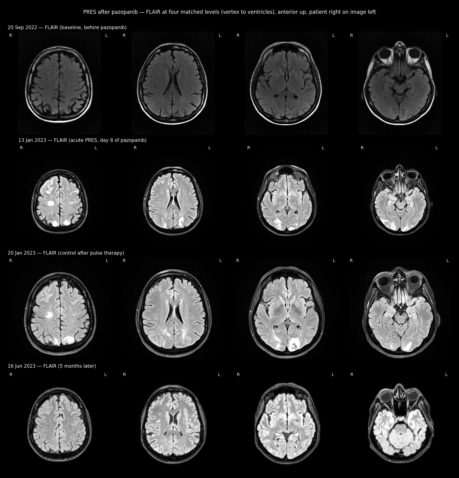
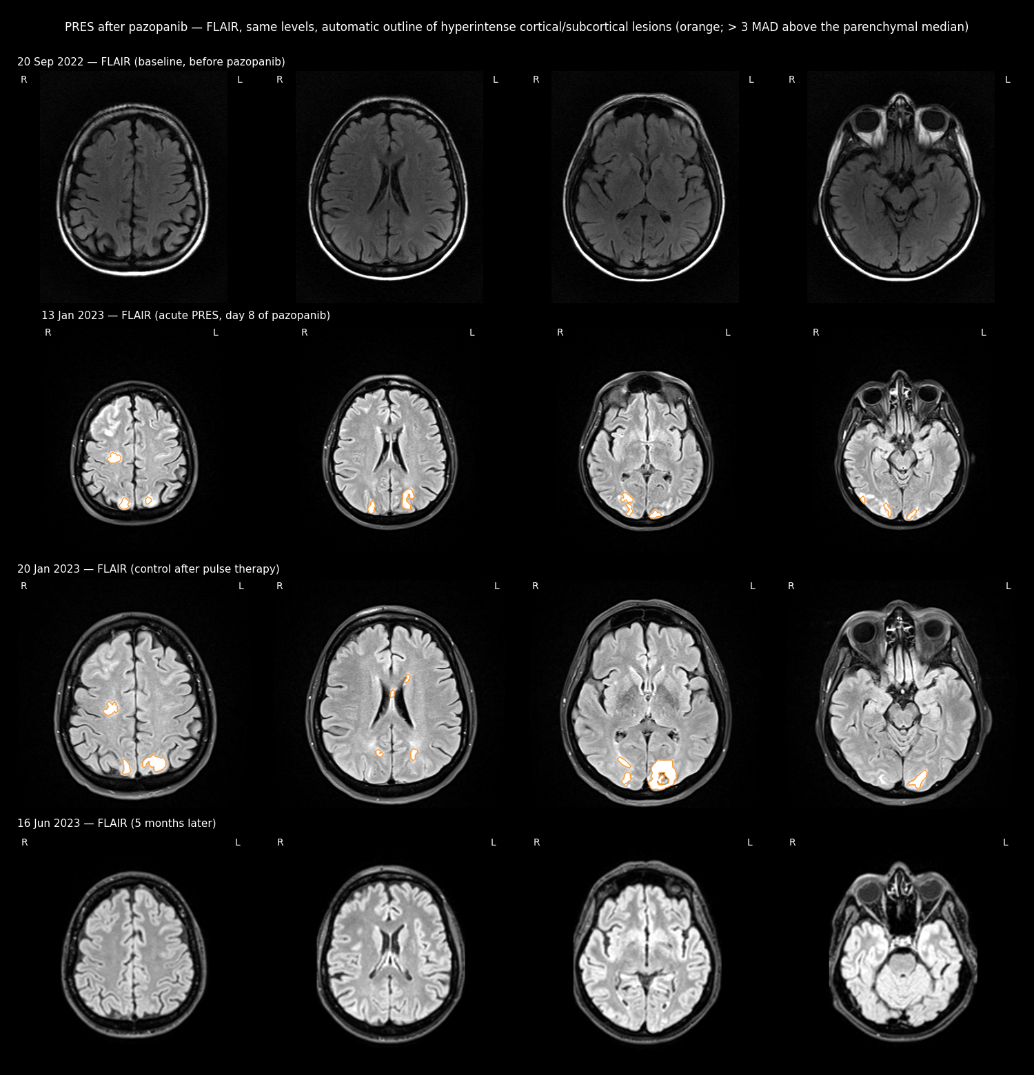
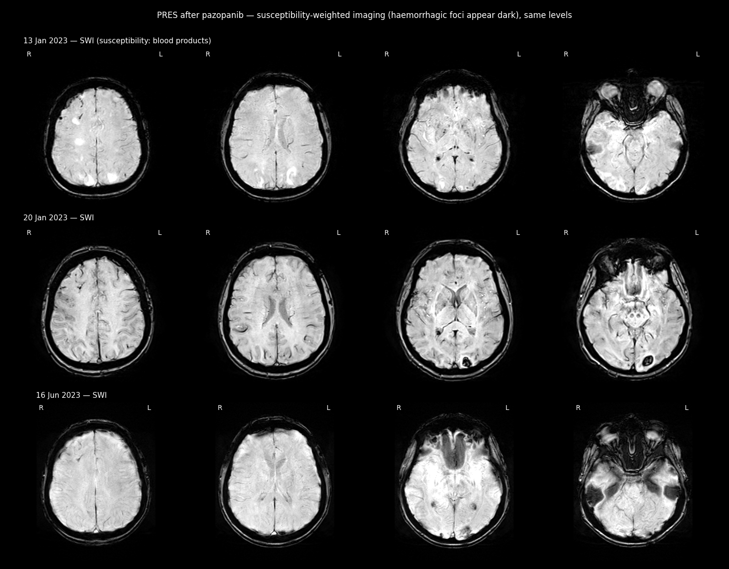
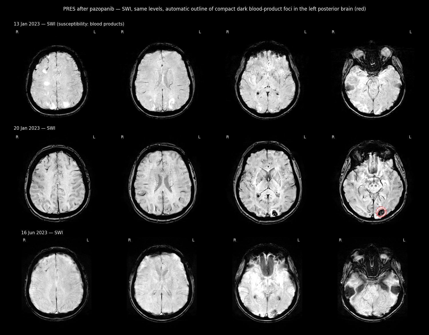
Imaging analysis of the 23.09.2026 PET-CT
Method. The radiologist's report and automatic organ maps were used only as starting points. Every structure was outlined slice by slice by AI segmentation agents and then checked by independent adversarial reviewer agents that re-rendered every slice and tried to refute each outline. Each outline was corrected until it passed review (all passed on the second review). Reference levels: liver background SUV 1.97 ± 0.63, FDG-active threshold 3.24 (liver mean + 2 SD), blood pool 1.58 (report: 1.6). Images use the radiological convention (patient right on the image left, anterior up).
| Structure | 23.09.2026 (own measurement) | 19.06.2026 | Radiologist 23.09.2026 |
|---|---|---|---|
| FDG-active tissue, left lobe (segments II/III) | 36.3 mL; 60 × 29 mm axial, 52 mm craniocaudal; SUVmax 6.66, 3.4 × liver; confluent with the lesion's medial side | 19 mL; SUVmax 2.27, 2.2 × liver | metabolic area 35 × 54 mm, confluent; SUVmax 4.9 (whole liver) |
| Left-lobe lesion, CT outline | 35 ± 4 mL; 45 × 30 mm axial, 56 mm craniocaudal | 27 mL; 46 × 48 × 42 mm | — |
| FDG-active focus, segment S8 (new) | 4.1 mL; 23 × 13 mm axial, 29 mm craniocaudal; SUVmax 6.75, 3.4 × liver; inside the hypodense upper lobule of the right-lobe mass | not active | 15 × 24 mm focus inside an ametabolic area |
| FDG-active wall of the right-lobe mass | 9.7 mL in 9 foci. The largest is the anterolateral plaque, 6.8 mL, SUVmax 6.02, 3.1 × liver; the medial spot next to the IVC is now 0.9 mL | 5.7 mL plaque (2.6 × liver) + 0.3 mL medial spot | "multiple foci of increased FDG" |
| Dominant right-lobe mass | 1200 mL; 146 × 116 mm axial, 180 mm craniocaudal; includes the 74 mL segment S8 lobule | ~1217 mL (same method) | 98 × 148 mm (the short axis is measured differently; our narrowest slices also give 98 mm) |
| Other liver lesions | none found (vessels and fissures excluded; lesions under ~8 mm cannot be excluded without contrast) | — | — |
| Liver | 2645 mL | — | — |
| Gastric-wall lesion (lesser curvature) | 36 mL; 42 × 31 mm, 52 mm craniocaudal; calcified, FDG-cold (core SUV ≈ 1.1) | 24 mL trace; 43 × 33 × 44 mm | 43 × 32 mm, calcified, FDG-negative |
| Anterior uterine lesion | 104 mL; 66 × 51 mm; SUV ≈ 1.0 × liver (maxima are bladder-urine spill-over) | — | 67 × 49 mm, SUVmax 1.8 |
| Upper-margin pelvic node | 23 mL; 43 × 37 × 29 mm (oblique); not FDG-active | — | ametabolic node 37 × 47 mm |
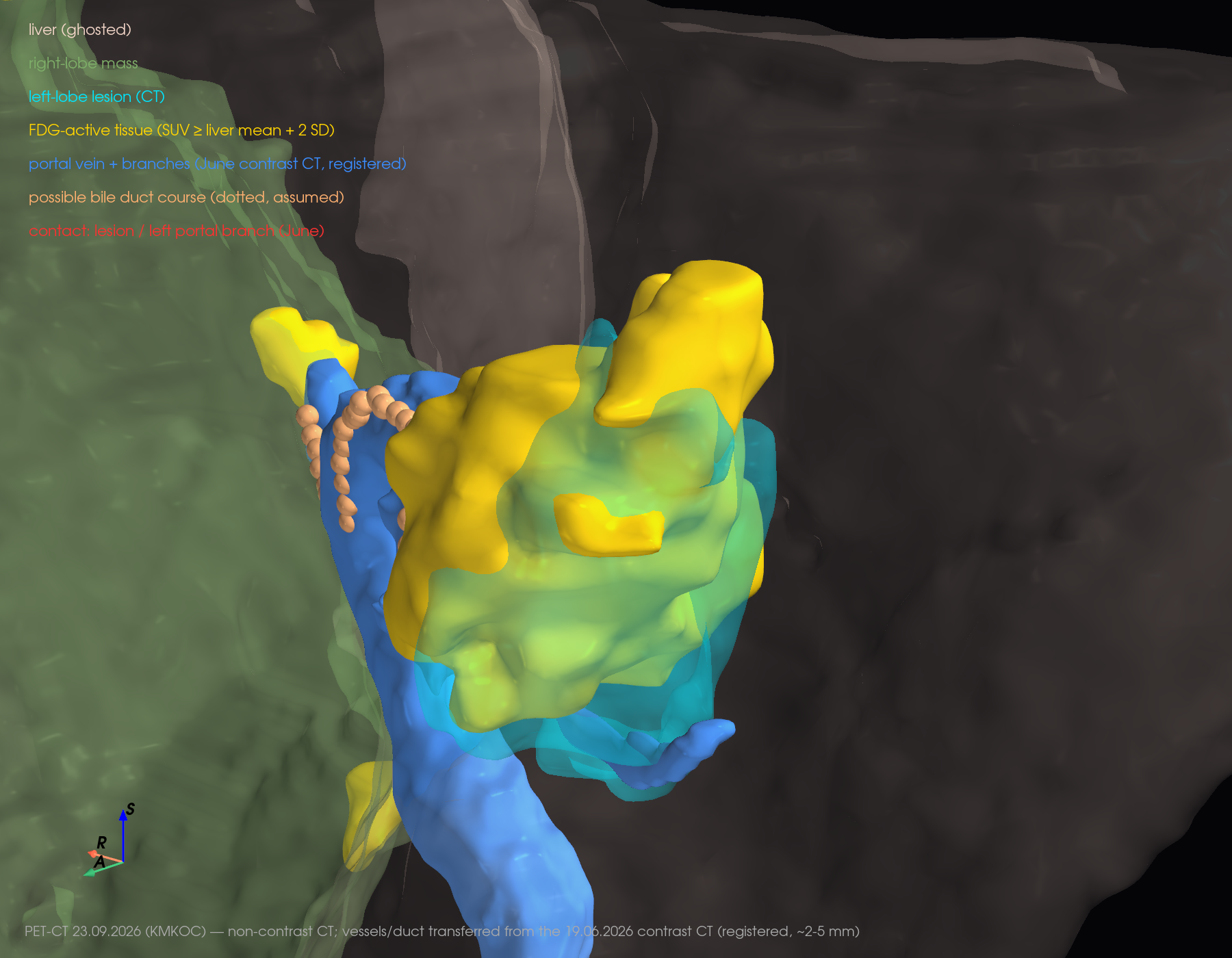
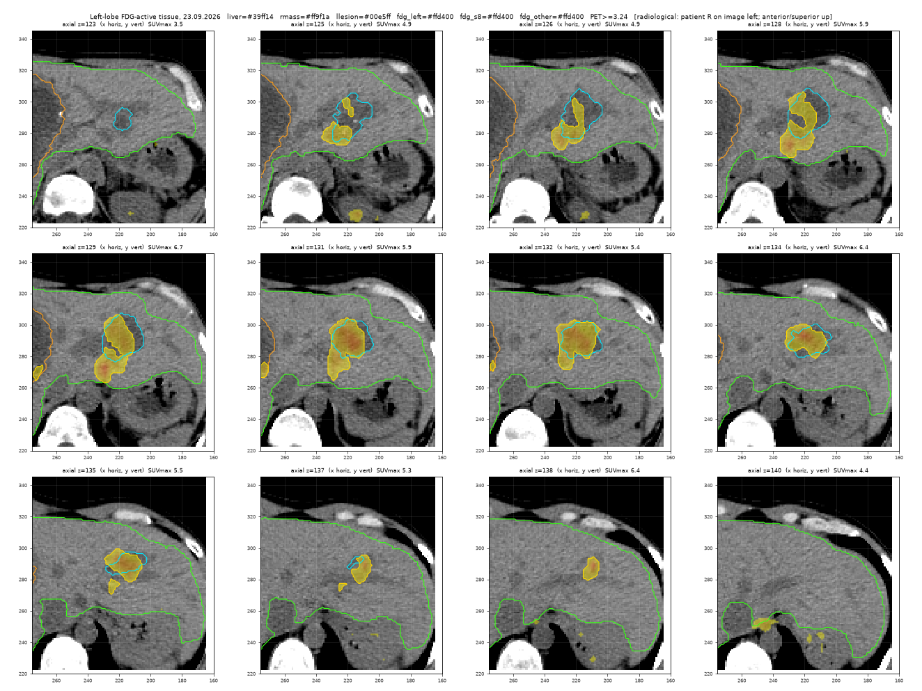
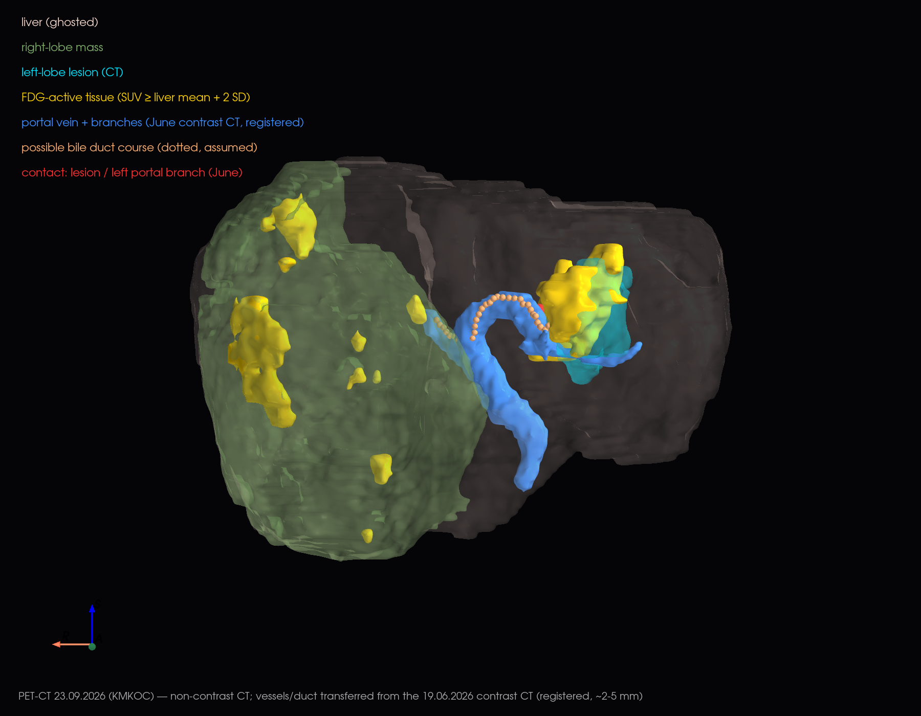
Segment S8 focus, right-lobe mass slices and other 3D views
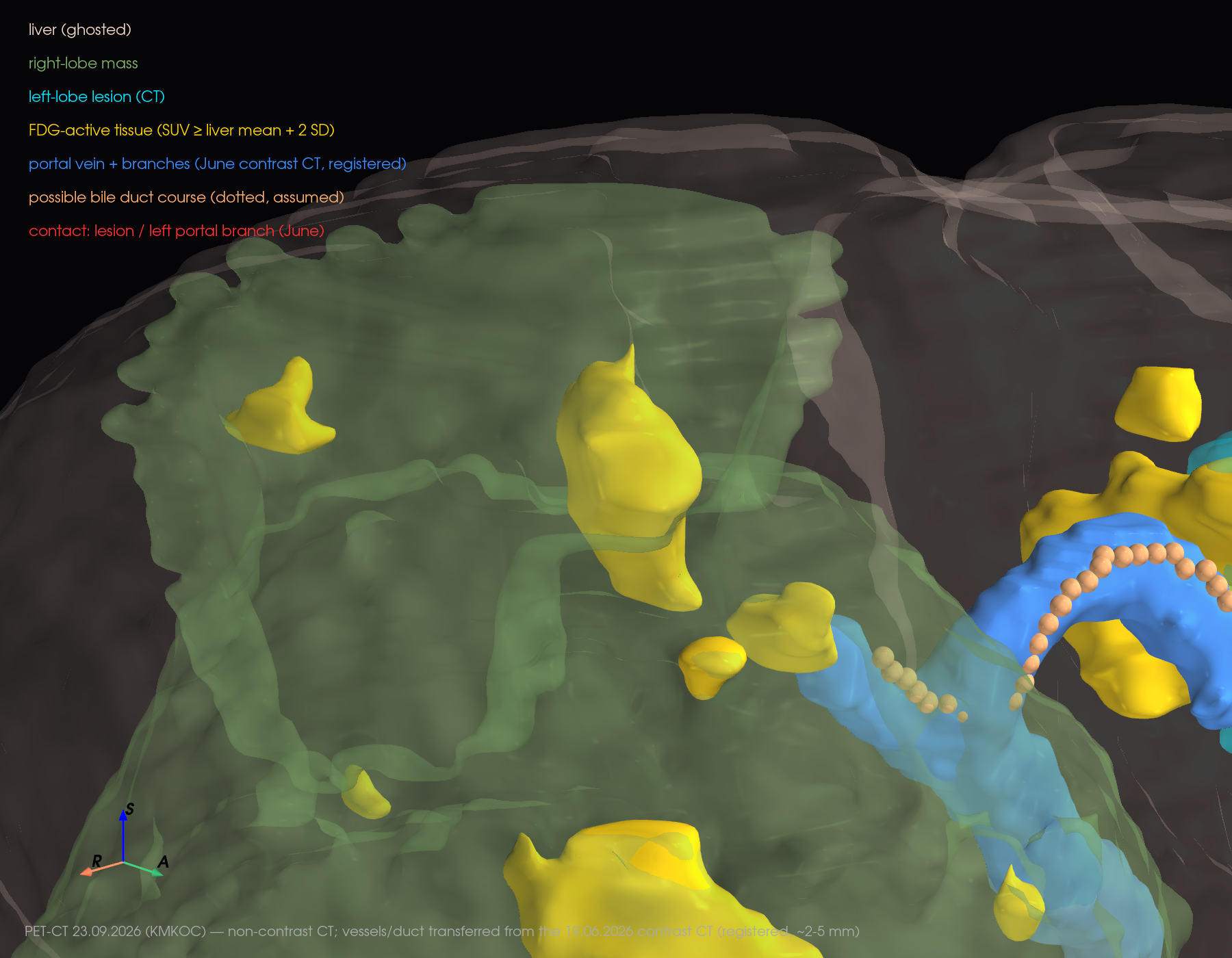
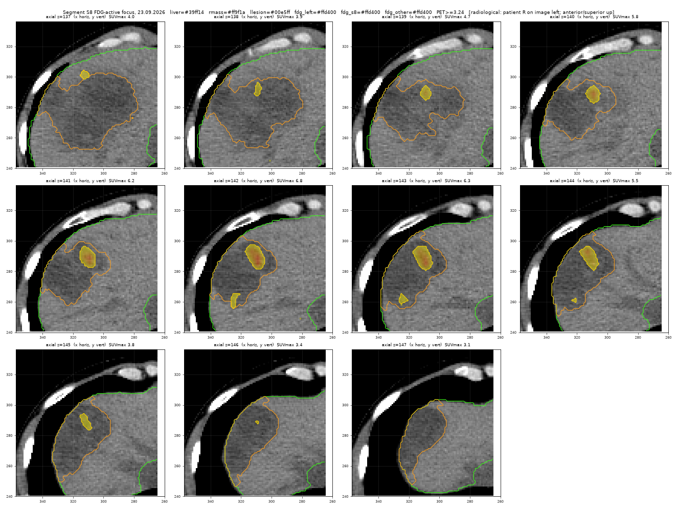
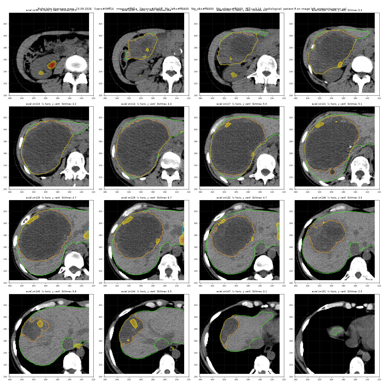
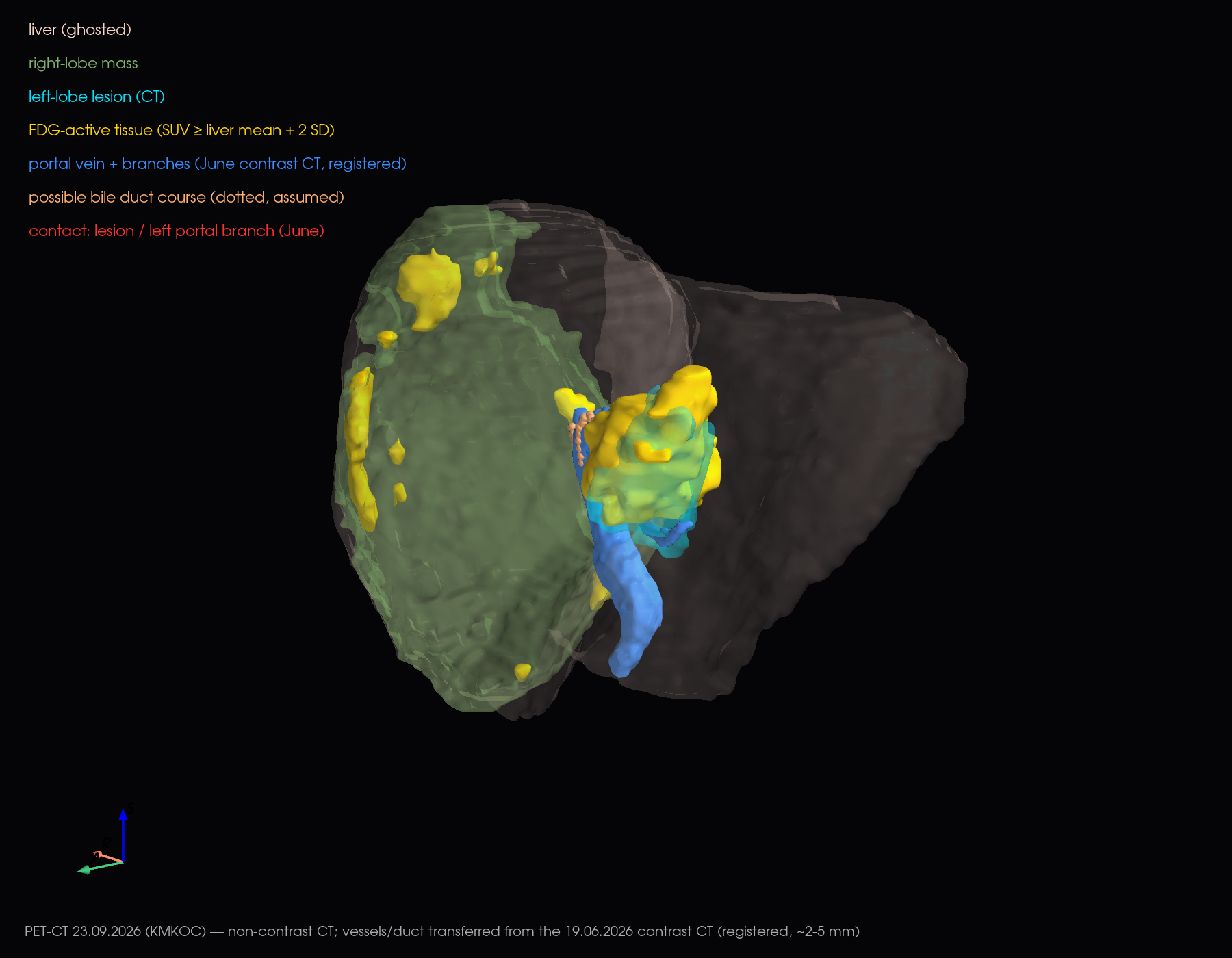
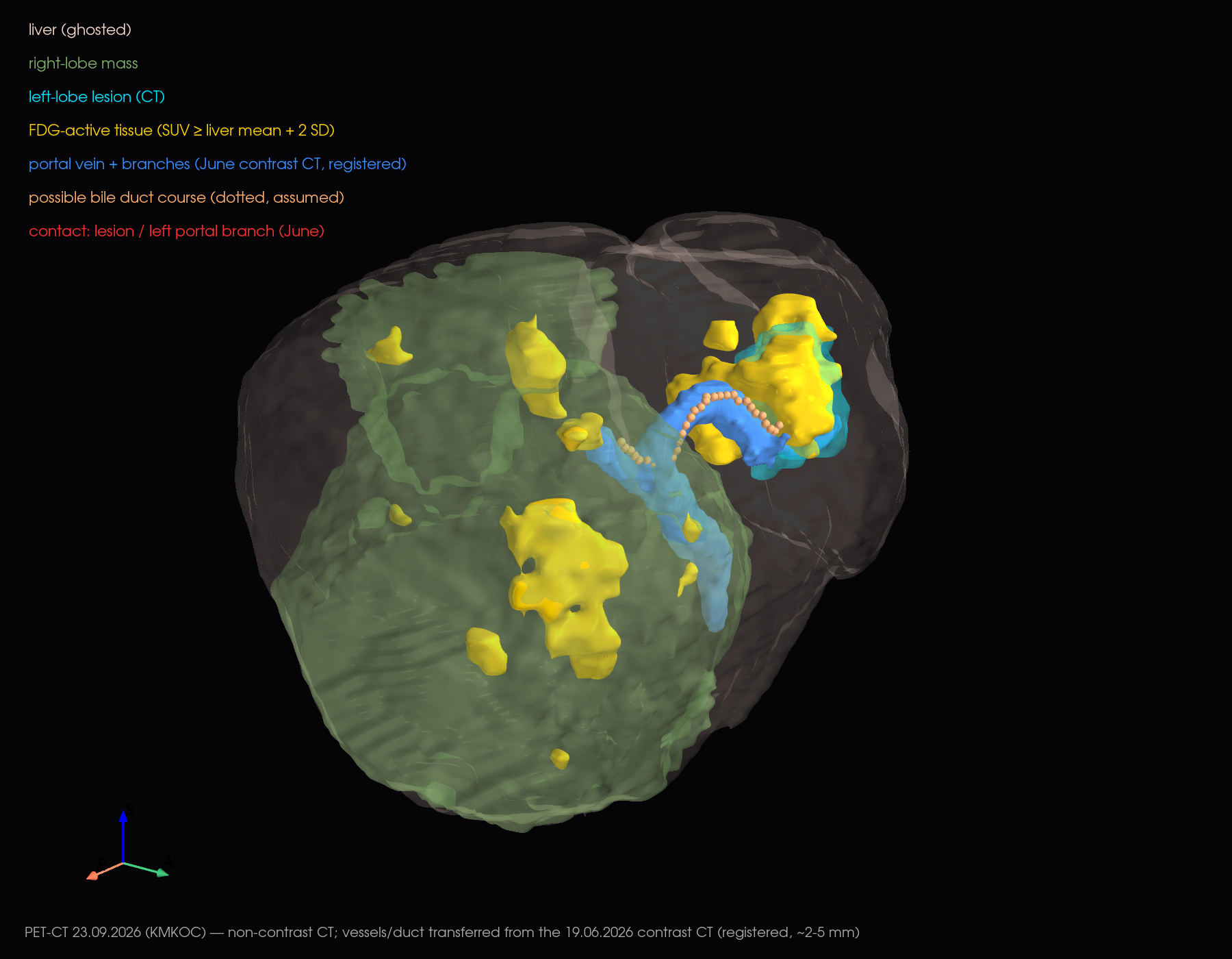
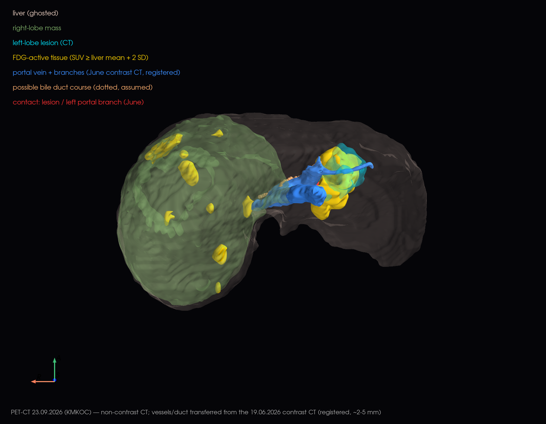
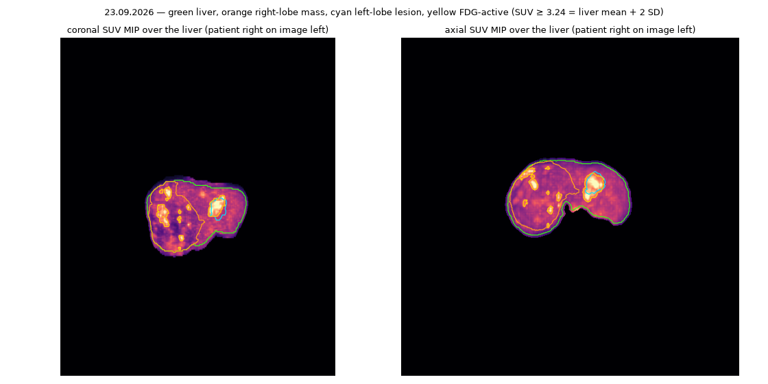
Gastric-wall lesion and pelvic lesions (slices)
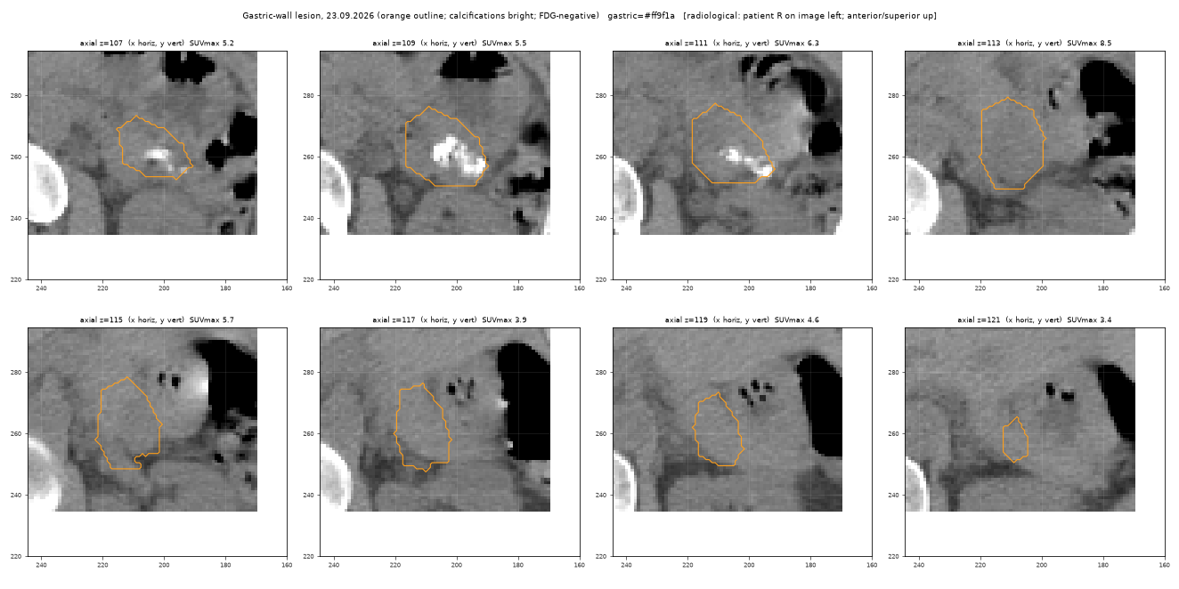
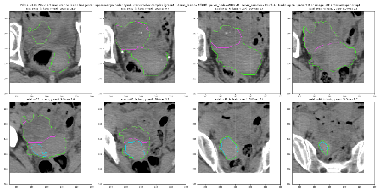
How exactly the 3D models match the slice segmentation (check)
Every 3D model is built directly from the reviewed slice masks, without pre-smoothing. Slices are interpolated linearly in the craniocaudal direction, the surface is placed half-way between an in-mask and an out-of-mask voxel, and only light volume-preserving smoothing is applied. Each model was checked by converting it back into voxels and comparing it with its mask. Agreement (Dice, 1 = identical) is 0.98–0.998, volumes agree within 0.5 %, and 95 % of each surface lies within about 1 mm of the voxel boundary (one voxel = 1.37 mm in-plane).
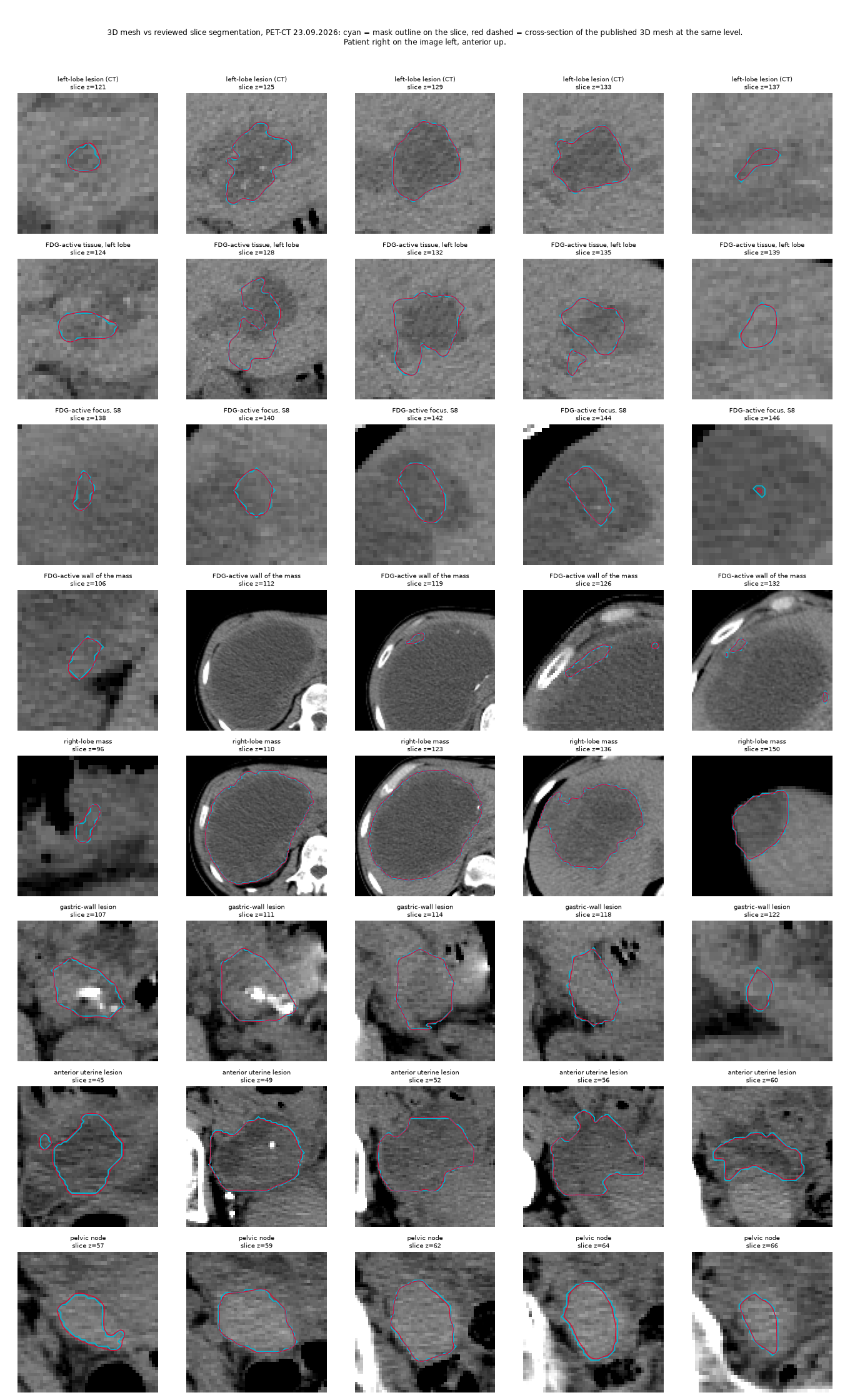
Portal vein and bile-duct course transferred from the June contrast CT (registration check)
The 23.09.2026 CT was done without iodine contrast, so the portal vein and its branches cannot be traced on it. They were traced on the 19.06.2026 contrast CT and transferred onto this study. The June liver, right-lobe mass and left-lobe lesion surfaces were aligned to the September ones (affine fit, then a local rigid refinement around the lesion). After alignment the June lesion surface lies on average 2.4 mm from the September lesion surface, and the liver surface within 60 mm of the lesion 5.3 mm from it. The transferred vessels are therefore approximate to a few millimetres, and they show the June anatomy. The bile duct is not visible on either study: its course is assumed from the portal branch it accompanies.
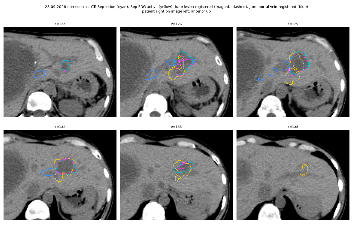
Imaging analysis of the 19.06.2026 PET-CT (10.09.2026)
The venous-phase contrast CT (2 mm) and the FDG PET of the 19.06.2026 study were fused using the DICOM geometry and reviewed slice by slice. Frame convention: RAS millimetres; images in radiological convention (anterior up, patient left on image right).
| Finding | Measurement |
|---|---|
| Left-lobe lesion, CT (hypodense, 44 HU vs liver 98 HU) | segment II (83 %) / III (17 %); 46 × 48 × 42 mm; 27 mL; centre RAS (−54, 173, −1201) |
| FDG-active tumour (SUVmax 2.27, liver background 1.04, threshold 1.65) | 19 mL; about half inside the hypodense core, the rest isodense enhancing tissue on the medial (hilar) side and inferior pole |
| Left portal branch | in contact with the lesion (0.8 mm) and with the FDG-active part (0 mm); contact point RAS (−35, 164, −1200) |
| Other structures | IVC 31 mm; stomach 19 mm; heart 28 mm; lesion reaches the liver surface (depth 0–31 mm) |
| FDG-active wall of the right-lobe mass (the "increased peripheral uptake" in the expert reading) | SUVmax 2.34 (2.6 × liver background 0.90; threshold 1.59 = liver mean + 2 SD); 5.7 mL plaque along the anterolateral wall, 27 × 24 × 54 mm, soft-tissue density (37 HU vs 15 HU cyst fluid), centre RAS (88, 195, −1220); plus a 0.3 mL spot (SUVmax 1.83) on the medial wall near the portal vein. No other part of the wall or cyst reaches the threshold. Blood-pool uptake is lower (IVC 0.94, portal vein 1.09), so neither spot is vascular spill-over. The intensity equals the left-lobe focus (2.27). |
| Bile ducts | not resolvable on CT (not dilated); the left hepatic duct runs in the same Glissonean sheath as the left portal branch, so it is within a few mm of the lesion |
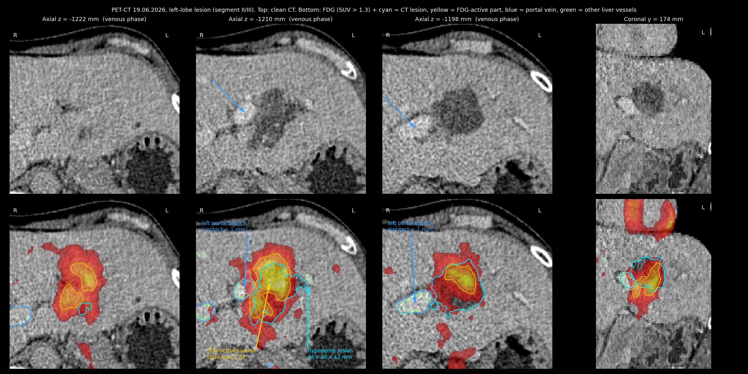
Nine consecutive axial slices through the lesion (Figure 2)
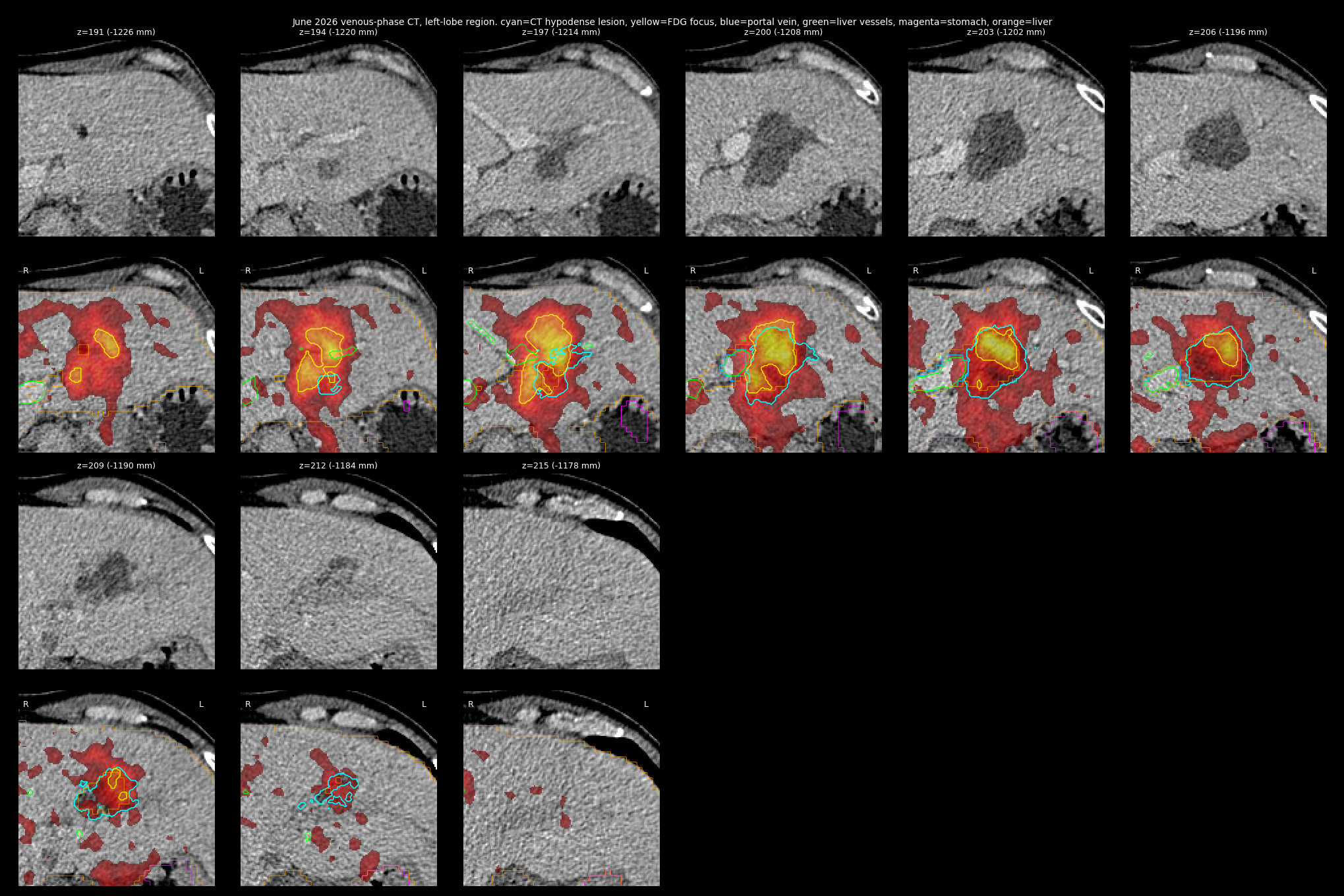
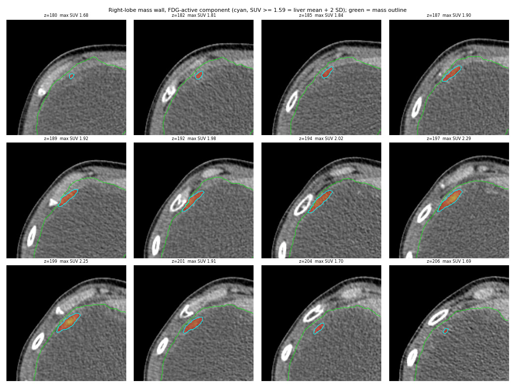
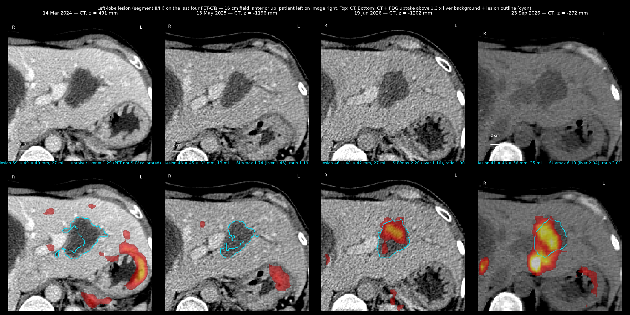
Dominant right-lobe mass on all six PET-CTs (Figure 4)
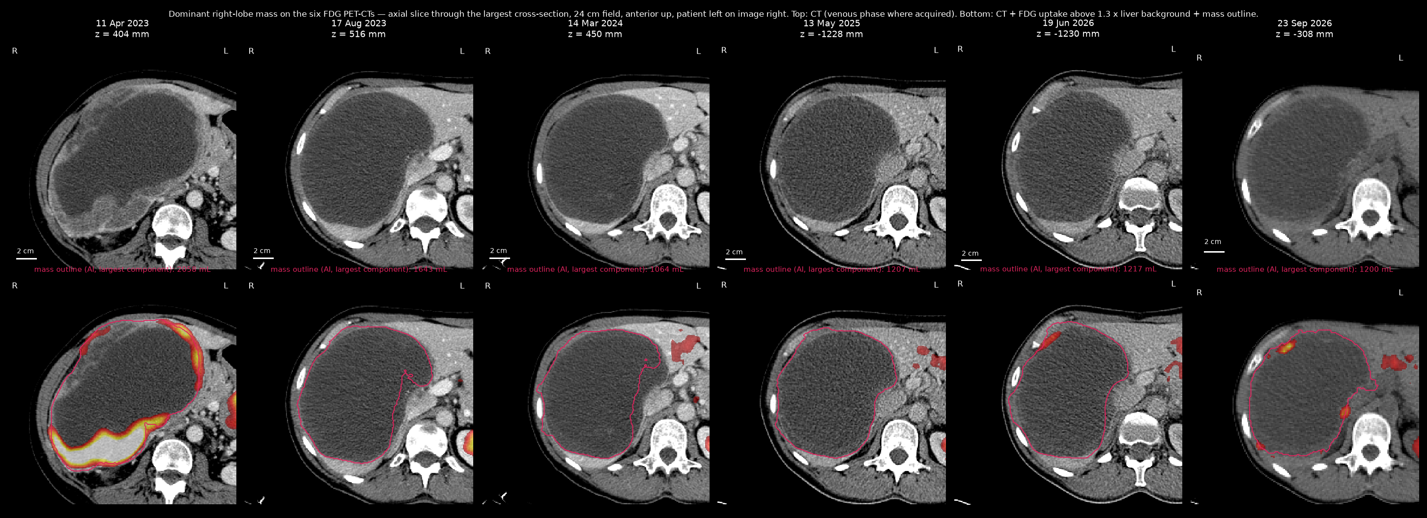
3D reconstruction
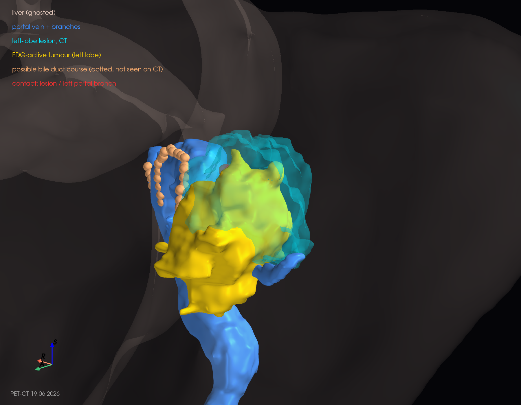
Slice-by-slice verification montages behind the 3D shapes
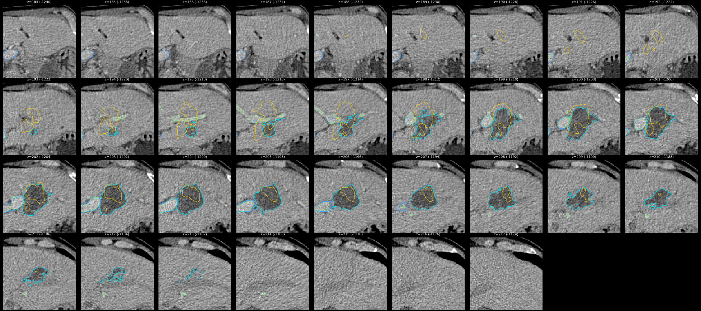
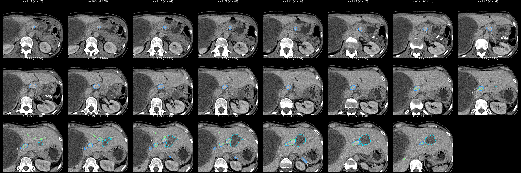
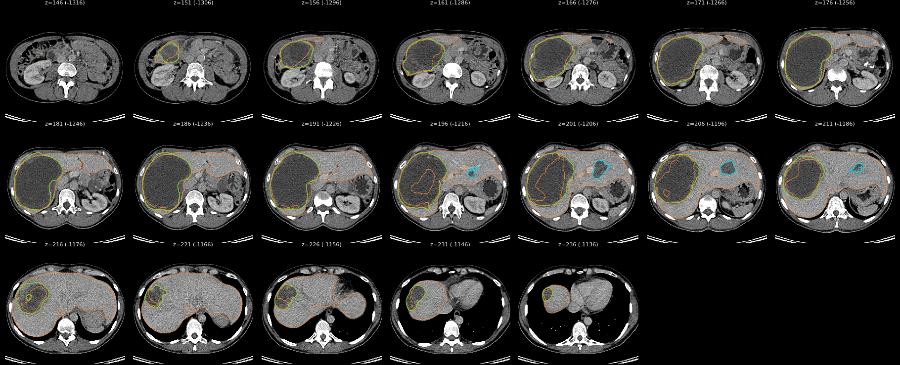
Gastric-wall GIST (eleven studies 2022–2026), uterus and right ovarian endometrioma (PET-CTs) over time
Method. On each study (gastric mass: five contrast CTs 2022–2023 from the DICOM discs plus six PET-CTs 2023–2026; pelvic masses: the six PET-CTs) the masses were traced slice by slice from a hand-placed seed (soft-tissue density window, air and bone excluded, growth limited to the tissue continuous with the seed), then every traced slice was checked visually (montages under the collapsible headings below). The traced masks give volume, the largest in-plane diameter and the maximum FDG uptake relative to the liver background. The 2023–2024 PET series were not exported in SUV units, so uptake is given relative to liver on all dates. The PET-CTs of 2023–2026 up to June 2026 were acquired with intravenous contrast. The 23.09.2026 study is a low-dose non-contrast CT, and its outlines come from the agent segmentation of that study that passed adversarial review (see the 23.09.2026 analysis), not from the seed tracer; its volumes are therefore not strictly comparable with the earlier traces (see the notes below each table).
Gastric-wall mass (primary GIST site), medial to the stomach at the level of the pancreatic body
| Study | Max in-plane diameter, mm | Volume, mL | Mean density, HU | Max FDG uptake / liver | Reported by the radiologists |
|---|---|---|---|---|---|
| 23 Aug 2022 (CT) | 55 | 53 | 63 | CT 23.08.2022: 51 × 46 mm above the pancreatic tail, adjacent to the stomach | |
| 1 Dec 2022 (CT) | 53 | 65 | 65 | CT 01.12.2022: 56 × 46 mm; German re-read: 5.2 × 6.1 cm | |
| 4 Jan 2023 (CT) | 52 | 70 | 58 | CT 04.01.2023: 65 × 50 mm, adjacent to the lower gastric body and the pancreatic-tail capsule | |
| 9 Feb 2023 (CT) | 50 | 52 | 51 | CT 09.02.2023: 55 × 40 mm (−15 % after pazopanib) | |
| 11 Apr 2023 | 54 | 36 | 75 | 5.5 × | 58 mm, SUVmax 8.2, active (PET-CT 11.04.2023) |
| 28 Jun 2023 (CT) | 54 | 47 | 43 | CT 28.06.2023: 47 × 35 mm, low density (−28 % on imatinib) | |
| 17 Aug 2023 | 48 | 46 | 41 | 1.8 × | 44 mm, almost fluid, no increased uptake |
| 14 Mar 2024 | 49 | 35 | 45 | 1.3 × | 40 mm, inactive |
| 13 May 2025 | 43 | 23 | 40 | 1.8 × | 35 mm, calcifications appearing |
| 19 Jun 2026 | 41 | 24 | 38 | 1.1 × | 43 × 33 × 44 mm (Feofaniya) / 37 × 31 mm, calcified, minimal uptake, unchanged (Israel) |
| 23 Sep 2026 | 41 | 36 | 51 | 1.2 × | 43 × 32 mm, calcified, FDG-negative, stable (KMKOC) |
Reading. The gastric-wall tumour grew through chemotherapy (53 → 65 → 70 mL from August 2022 to January 2023, enhancing, 58–65 HU), shrank after the week of pazopanib (52 mL in February 2023) and was intensely FDG-active in April 2023; on imatinib it turned cystic (47 mL and 43 HU by June 2023, almost fluid by August 2023) and then settled at 23–35 mL (about 4 cm) through 2024–2026 with progressive calcification, mean density around 40 HU and uptake at or near liver background. This matches the reports (unchanged, calcified, minimal uptake). Note on April 2023: the enhancing, intensely active mass at the lower gastric body next to the pancreatic tail was traced from its FDG-avid volume (restricted to soft-tissue density), because on CT its enhancement matched the adjacent tissue; the result (about 5 cm, uptake about 5.5 × liver) agrees with the report (58 mm, SUVmax 8.2), but its volume (36 mL) counts only the FDG-avid part and is therefore lower than the CT-defined volumes of the neighbouring dates. The 75 mm left-lobe liver metastasis, which lay 10 cm higher under the diaphragm that month, is a separate object. 23.09.2026: unchanged in linear size (41 mm, as in June) and FDG-cold. The volume of 36 mL against 24 mL in June reflects the method, not growth: the September outline includes the calcified lateral clumps that the June trace left outside.
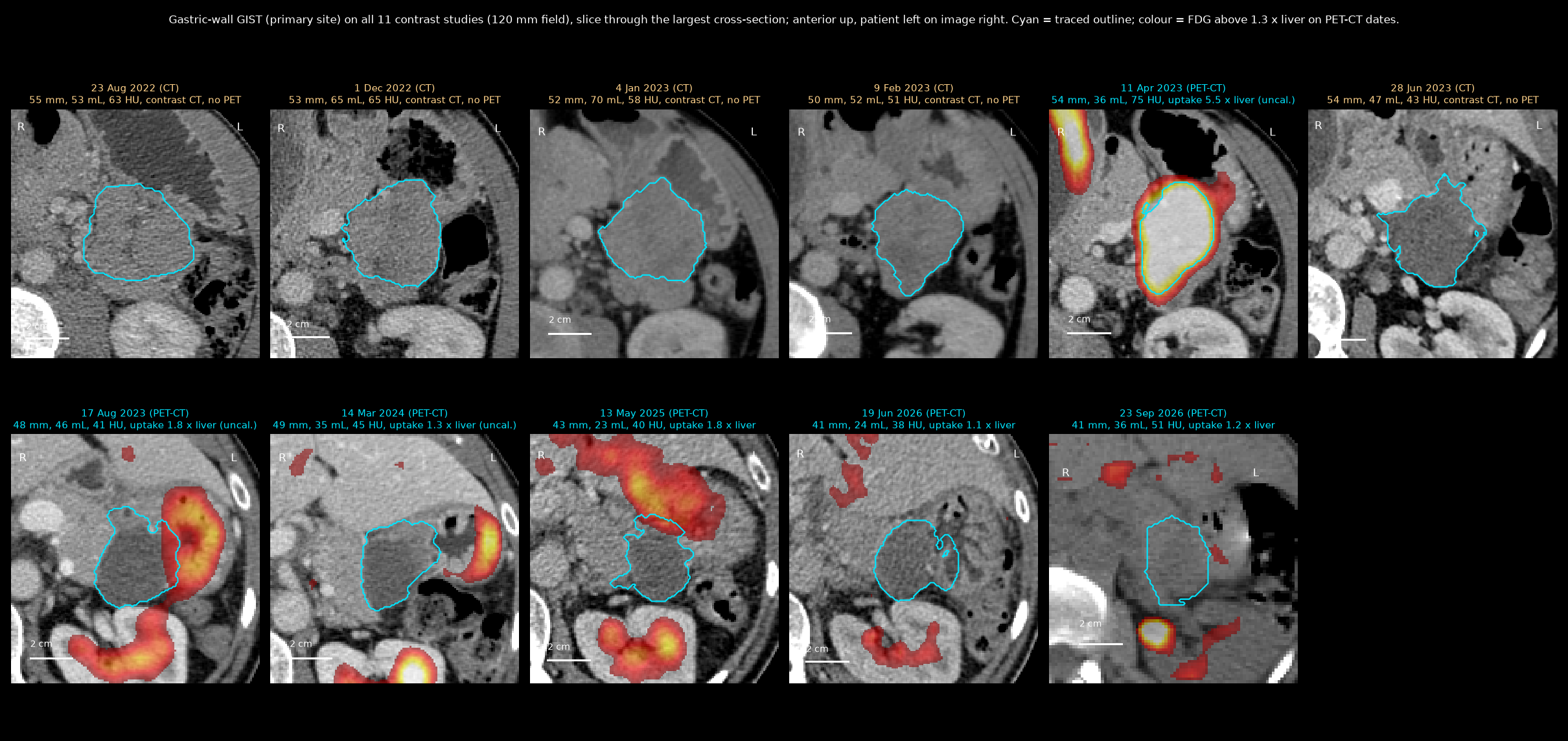
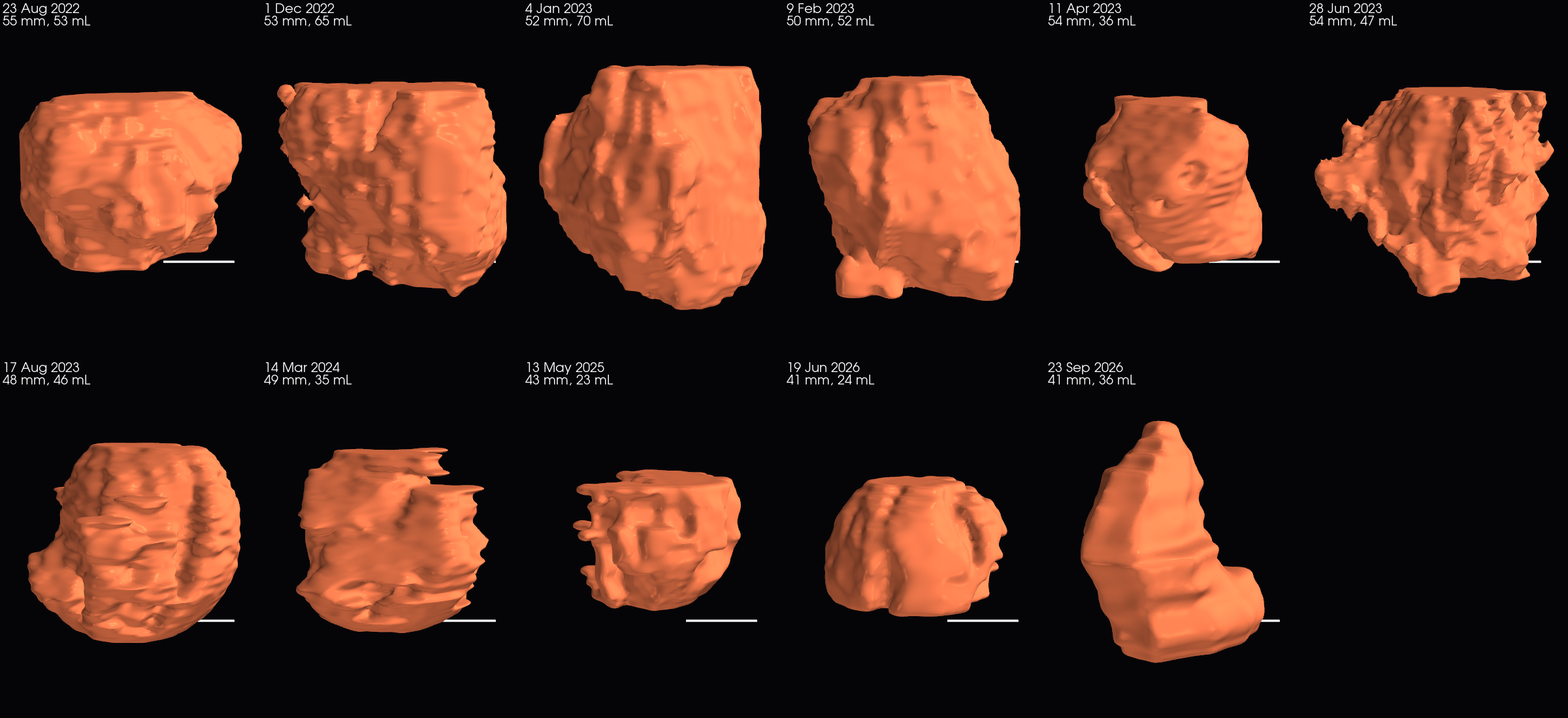
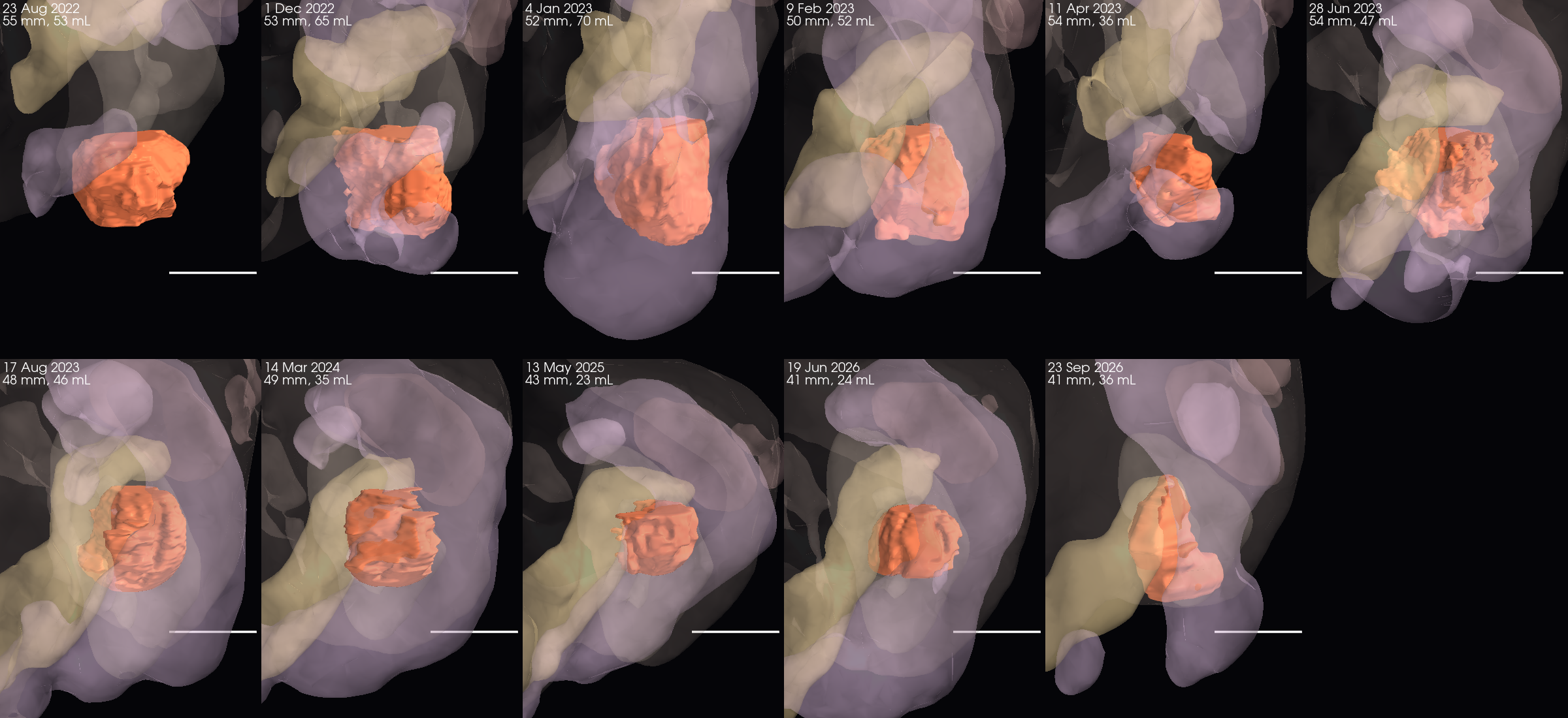
Uterus with leiomyomas — the whole organ traced on the six PET-CTs (uterine body plus fibroids)
| PET-CT | Max in-plane diameter, mm | Volume, mL | Mean density, HU | Max FDG uptake / liver | Reported by the radiologists |
|---|---|---|---|---|---|
| 11 Apr 2023 | 110 | 107 | 73 | 1.8 × | inactive uterine mass 72 mm |
| 17 Aug 2023 | 101 | 99 | 76 | 1.5 × | 96 mm, inactive |
| 14 Mar 2024 | 98 | 100 | 75 | 1.3 × | 78 mm, smaller |
| 13 May 2025 | 108 | 134 | 55 | 1.6 × | 84 mm |
| 19 Jun 2026 | 92 | 153 | 51 | 2.4 × | 68 × 55 × 48 mm plus upper-margin mass 46 × 37 mm, stable, low activity (Feofaniya); heterogeneous uterine lesions without dynamics (Israel) |
| 23 Sep 2026 | 113 | 248 | 31 | 2.2 × | anterior-wall lesion 67 × 49 mm, SUVmax 1.8, and ametabolic upper-margin node 37 × 47 mm, both stable (KMKOC) |
Reading. The violet object is the whole uterus (body plus several fibroids, traced as one organ because fibroid and myometrium cannot be separated reliably on CT); this is why it is larger than the single-fibroid diameters in the reports. On the 2022–2023 contrast CTs only the dominant homogeneous pelvic mass could be traced consistently, so those dates are not included in this series. It stays between 130 and 190 mL with no consistent trend over three years and low uptake (up to 1.5–1.8 × liver; the 2.4 × in June 2026 comes from a bowel loop touching the outline). This is the behaviour of leiomyoma, not of the GIST metastases, and it agrees with the reports ("without dynamics"). Our numbers are larger than the single-mass diameters in the reports because the whole uterine complex is traced; on some dates (notably August 2023, patient's left side) the outline may include an adjacent bowel loop. 23.09.2026: the 248 mL is mostly a difference of method, not growth. The September outline reaches lower, to the bladder dome, and includes the anterior lesion that the June trace cut off at its lower edge; the June outline was also ragged and under-inclusive. Compared over the same levels the difference is between −4 % (above the bladder dome: 147 vs 153 mL) and +41 % (a 60 mm block aligned on the node), within the 134–228 mL spread of the earlier traces. The radiologist reports both pelvic lesions as stable.
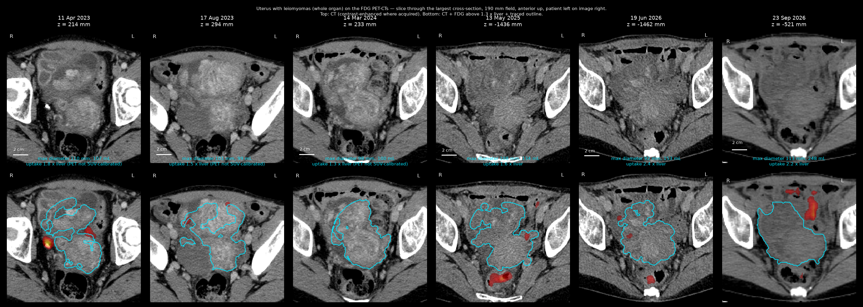
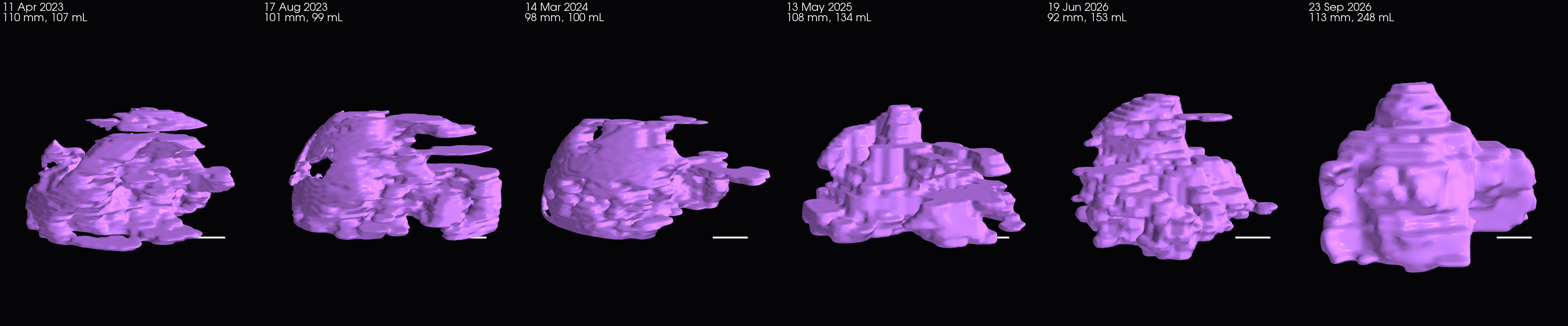
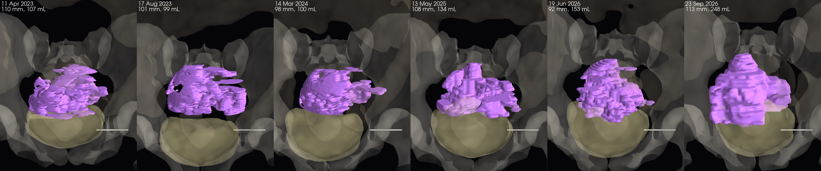
Right ovarian endometrioma (haemorrhagic cyst of the right ovary; "endometrioma" per the 2023 Israeli MRI/CT revision)
A homogeneous, non-enhancing (about 45 HU), smoothly outlined mass in the projection of the right ovary / pouch of Douglas, present since the first CT of August 2022 (67 × 57 mm). It was separated from the uterus trace as the low-density component inside it (below 55–62 HU after smoothing); on the 2025 and 2026 studies the whole lower uterus is of similar density and no distinct component could be isolated, in line with the 2026 report ("ovaries not clearly differentiated").
| PET-CT | Max in-plane diameter, mm | Volume, mL | Mean density, HU | Max FDG uptake / liver | Reported by the radiologists |
|---|---|---|---|---|---|
| 11 Apr 2023 | 82 | 64 | 45 | 1.3 × | about 6 cm (Israeli revision 19.04.2023: ~6 cm, ~50 HU, no enhancement); CT 09.02.2023: 75 × 45 mm, +45 HU |
| 17 Aug 2023 | 84 | 84 | 39 | 1.5 × | CT 28.06.2023: 65 × 42 mm, +45 HU, clear even contours |
| 14 Mar 2024 | 80 | 44 | 43 | 1.2 × | Israeli revision 25.03.2024: caudal formation 4.8 × 5.1 cm (from 5 × 5.5) |
| 13 May 2025 | not separable from the uterus on this study: no distinct non-enhancing right-adnexal component (the whole lower uterus is of similar density) | not described separately ("ovaries not clearly differentiated" in 2026) | |||
| 19 Jun 2026 | not separable from the uterus on this study: no distinct non-enhancing right-adnexal component (the whole lower uterus is of similar density) | not described separately; pelvic MRI recommended | |||
| 23 Sep 2026 | not separable on this non-contrast study (two candidate regions inside the pelvic complex) | not described separately (KMKOC) | |||
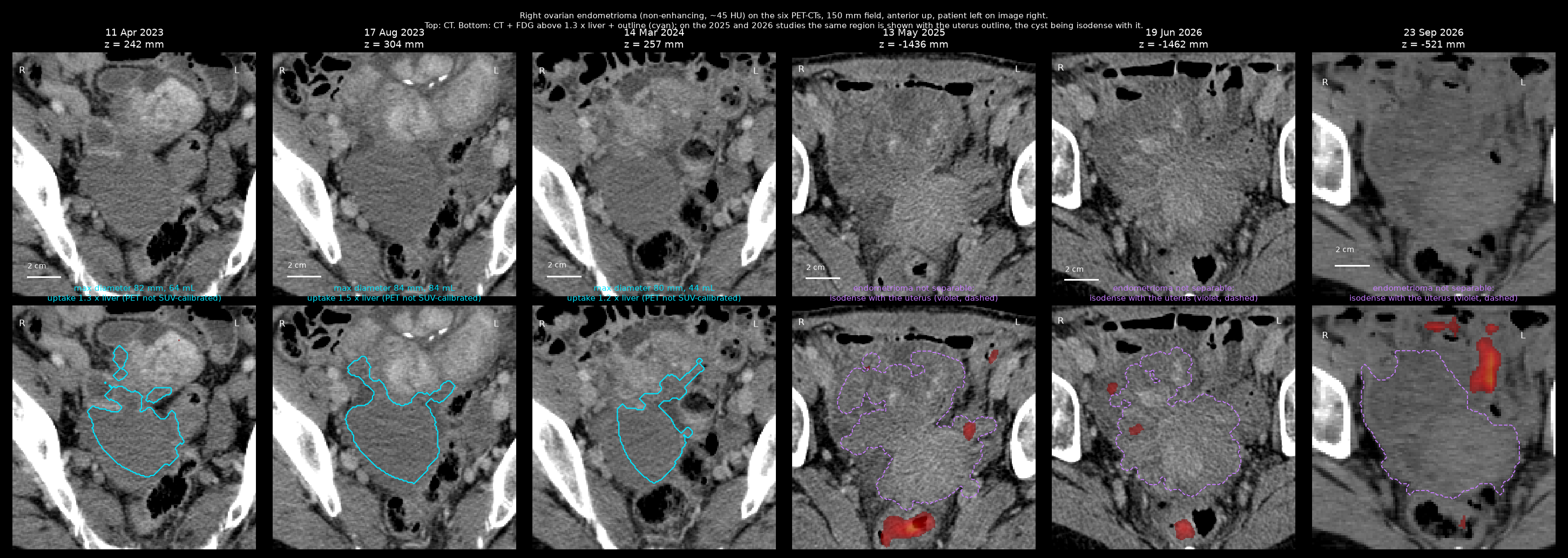
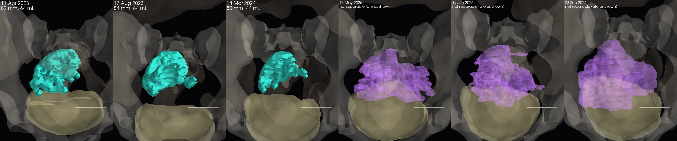
Slice-by-slice verification montages — gastric-wall mass (eleven dates)
23 Aug 2022 (contrast CT) — every traced slice (gastric)
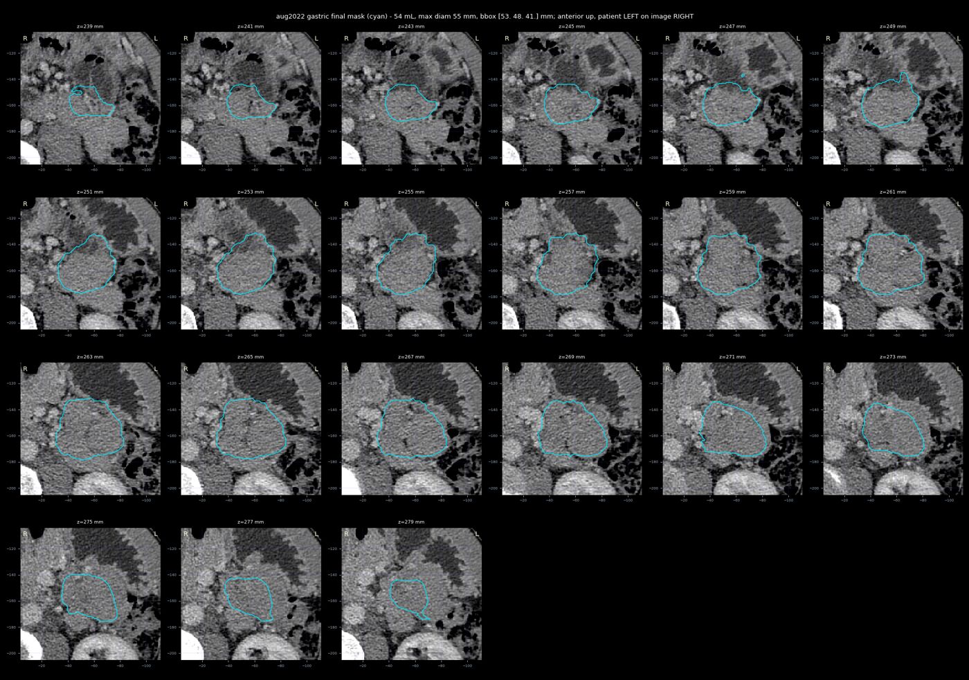
1 Dec 2022 (contrast CT) — every traced slice (gastric)
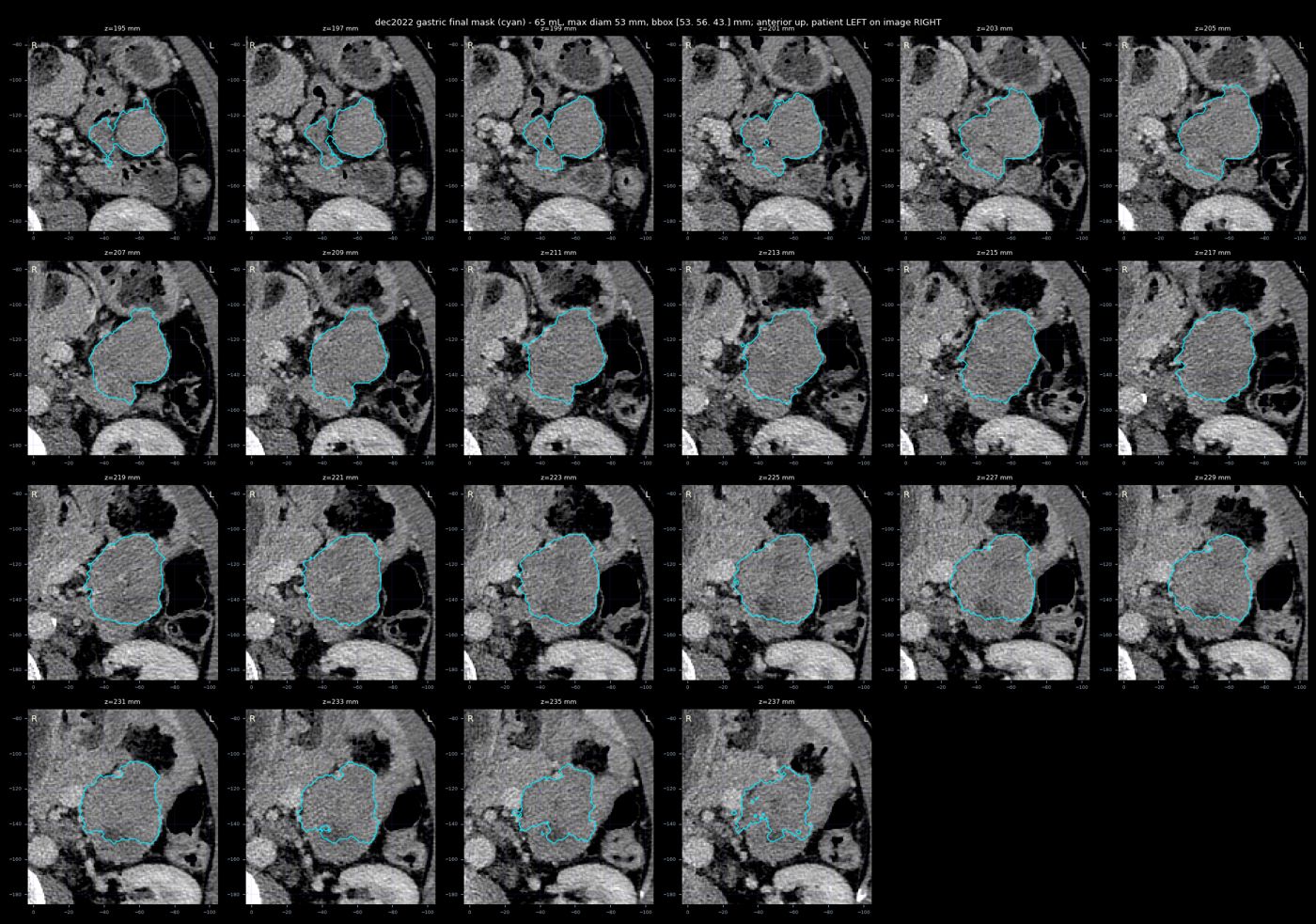
4 Jan 2023 (contrast CT) — every traced slice (gastric)
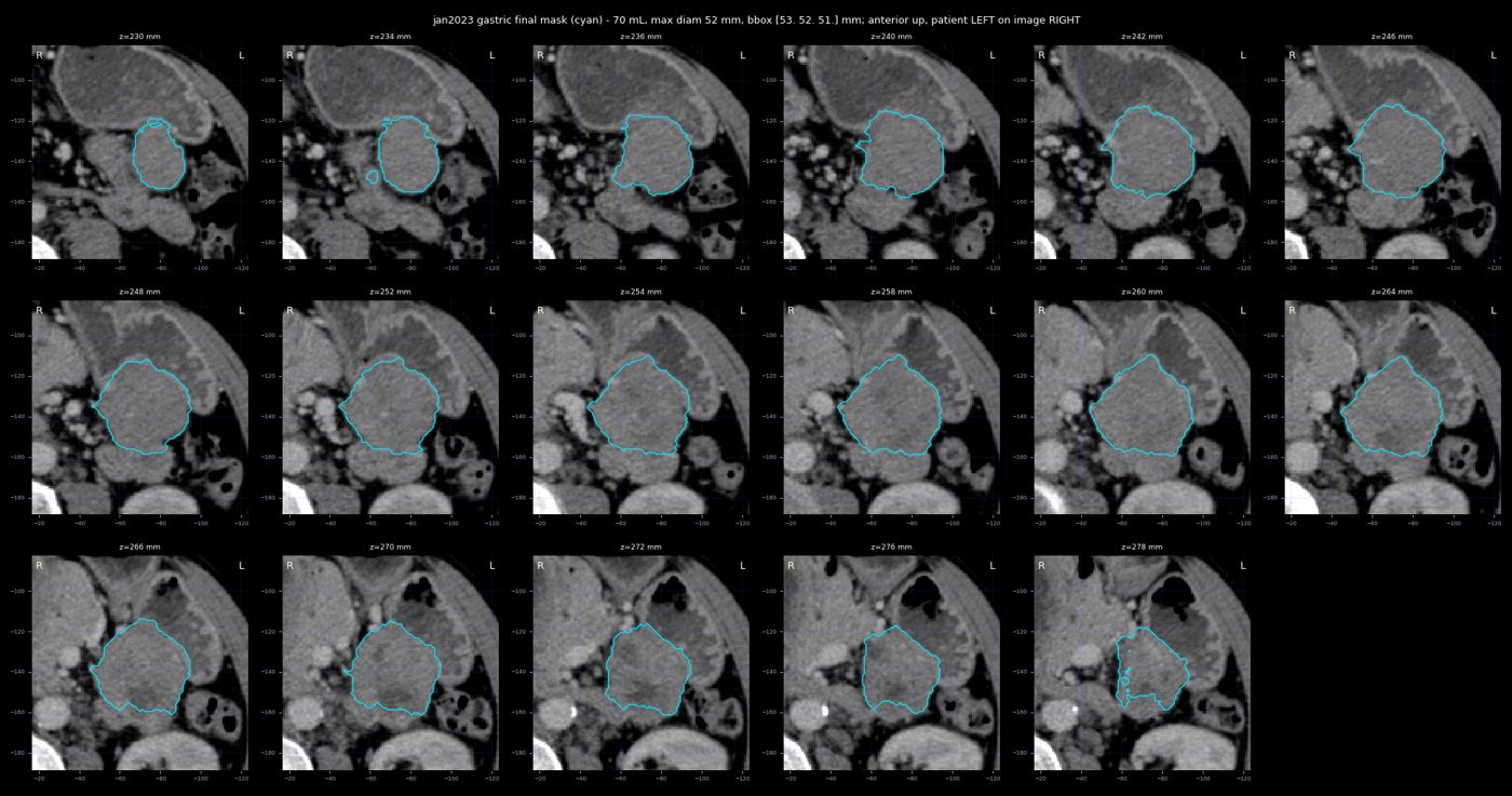
9 Feb 2023 (contrast CT) — every traced slice (gastric)
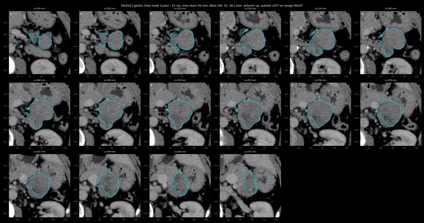
28 Jun 2023 (contrast CT) — every traced slice (gastric)
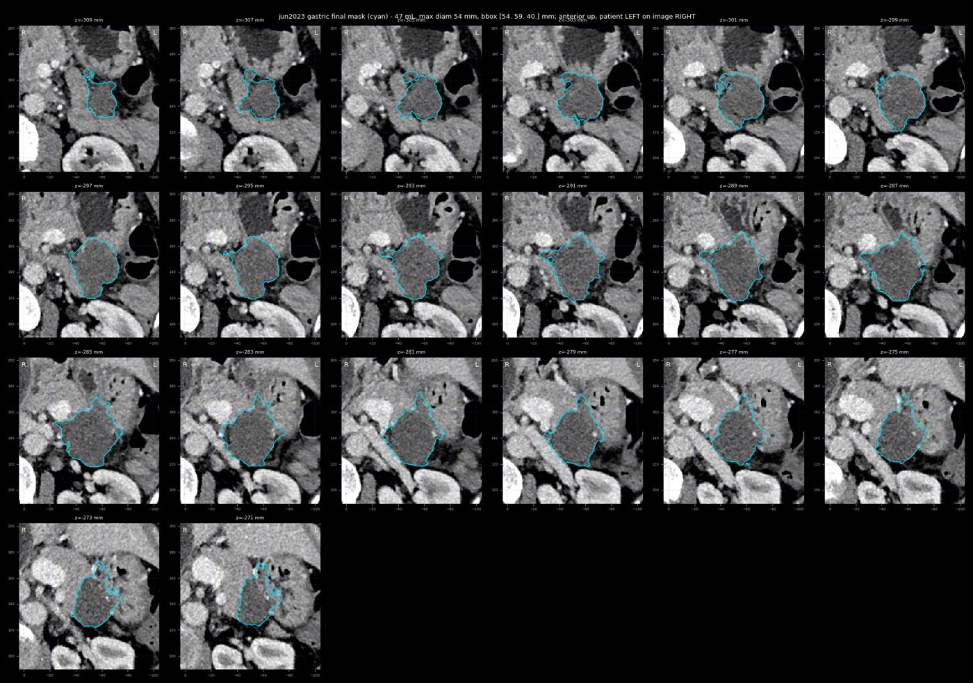
11 Apr 2023 — every traced slice (gastric)
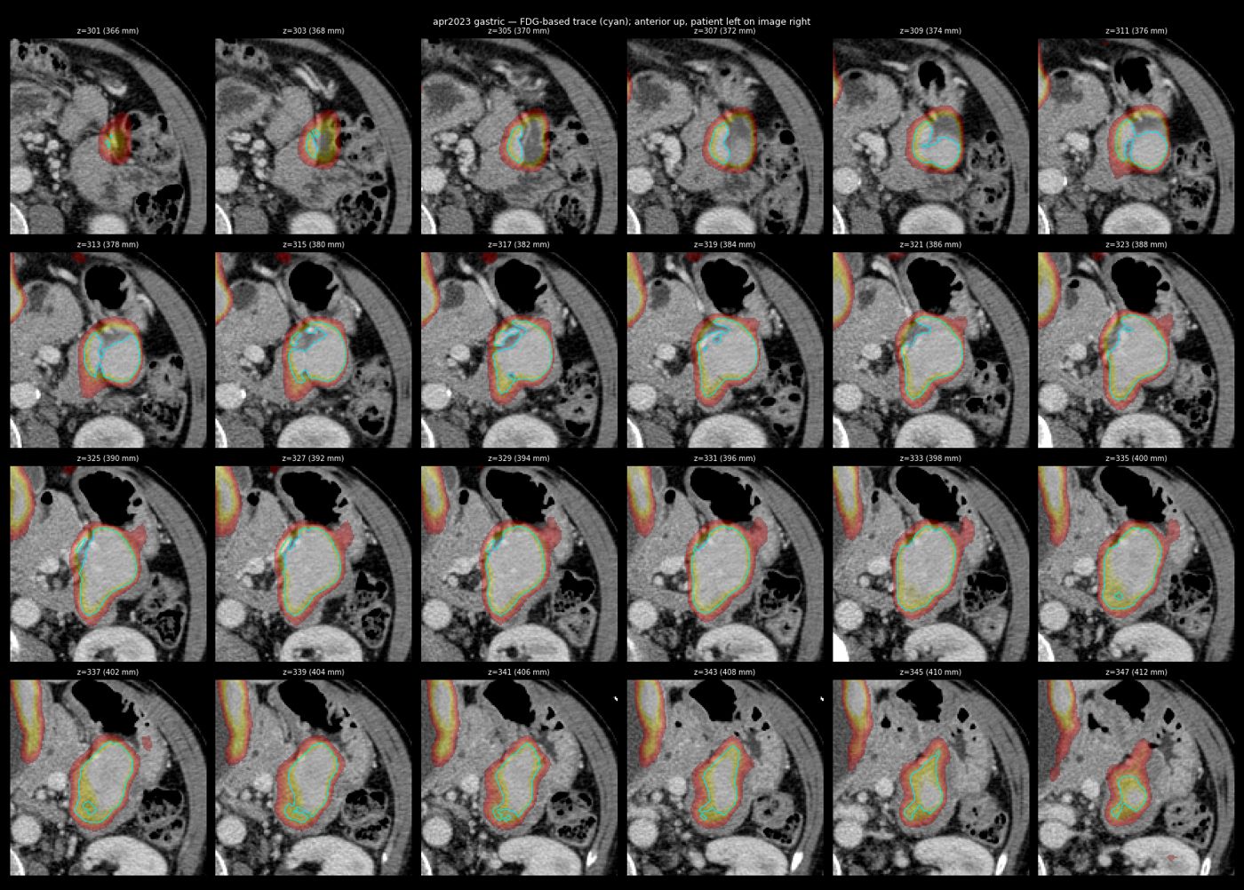
17 Aug 2023 — every traced slice (gastric)
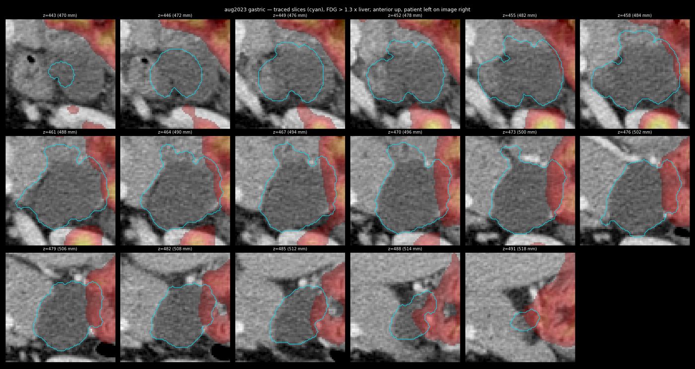
14 Mar 2024 — every traced slice (gastric)
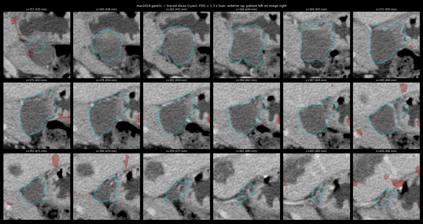
13 May 2025 — every traced slice (gastric)
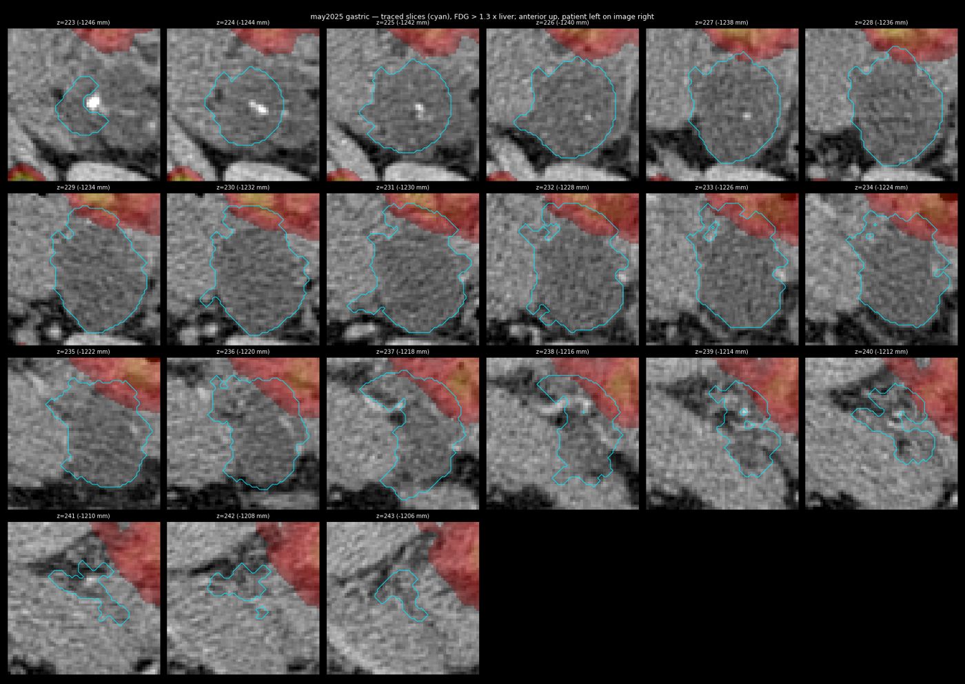
19 Jun 2026 — every traced slice (gastric)
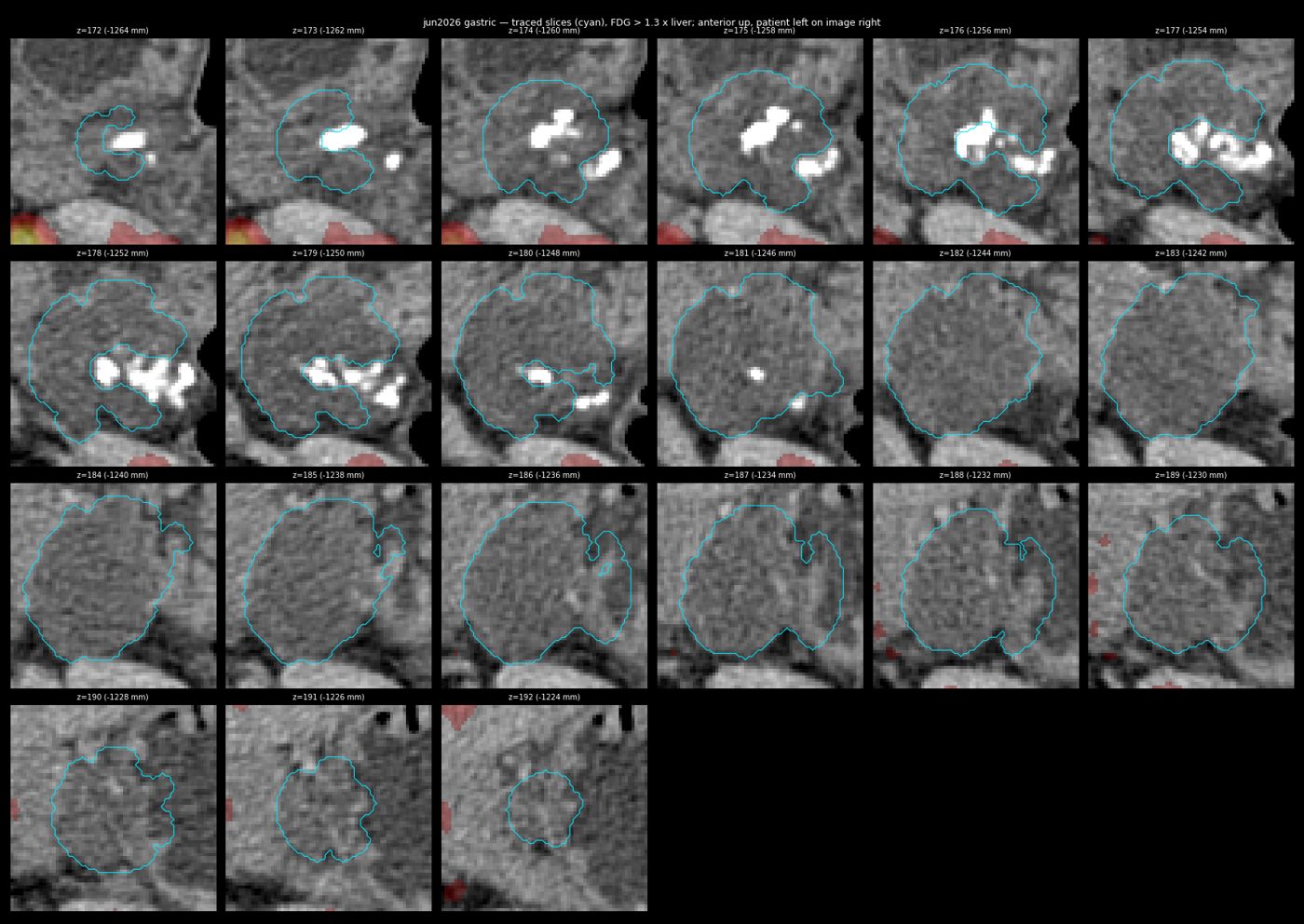
23 Sep 2026 (non-contrast; reviewed agent segmentation) — every traced slice (gastric)
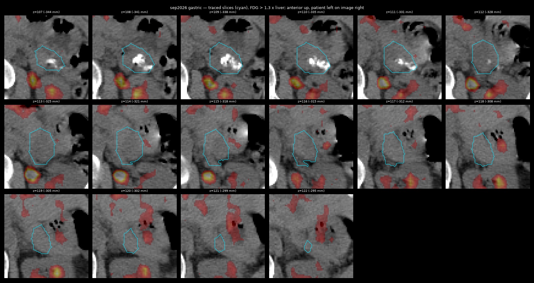
Slice-by-slice verification montages — uterus (six PET-CT dates)
11 Apr 2023 — every traced slice (uterus)
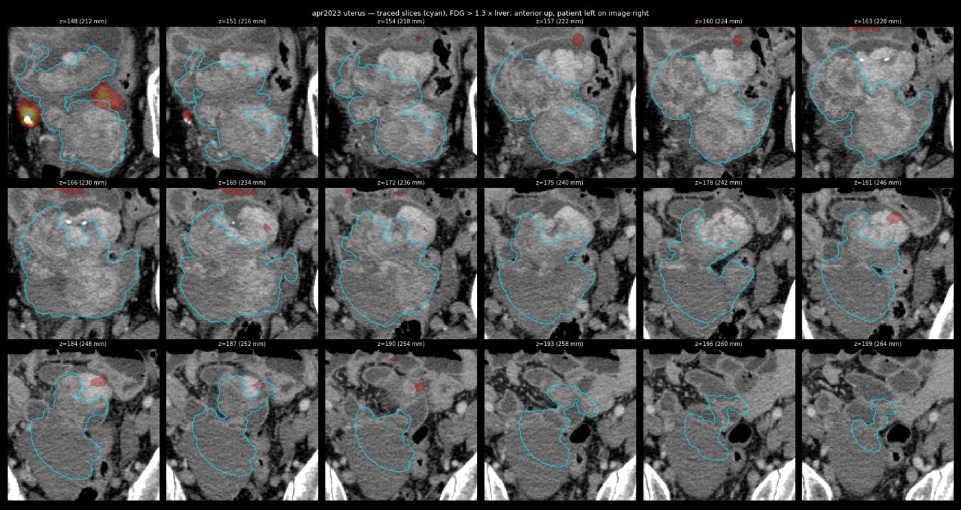
17 Aug 2023 — every traced slice (uterus)
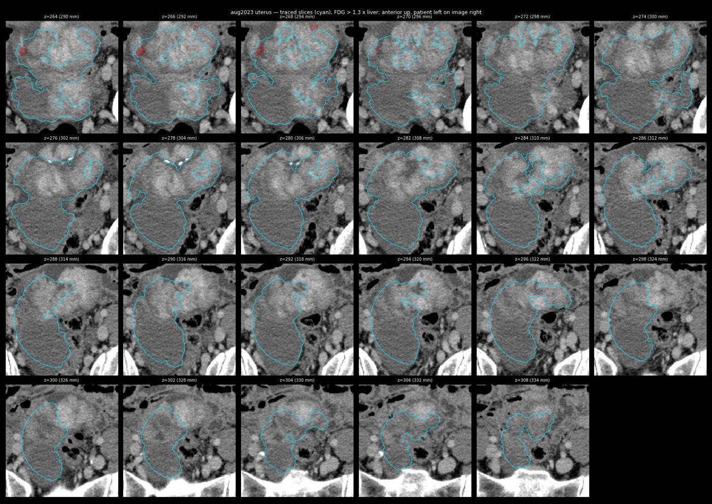
14 Mar 2024 — every traced slice (uterus)
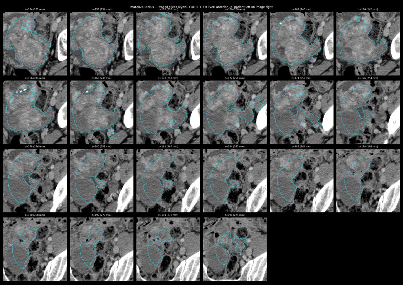
13 May 2025 — every traced slice (uterus)
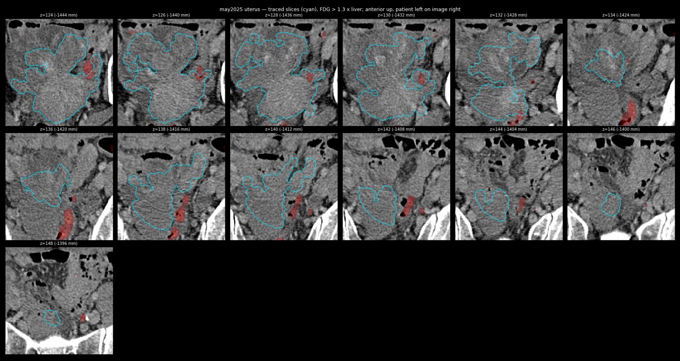
19 Jun 2026 — every traced slice (uterus)
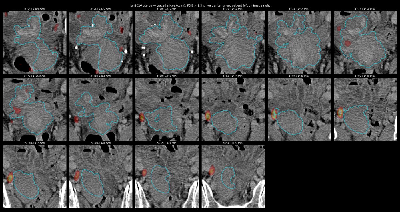
23 Sep 2026 (non-contrast; reviewed agent segmentation) — every traced slice (uterus)
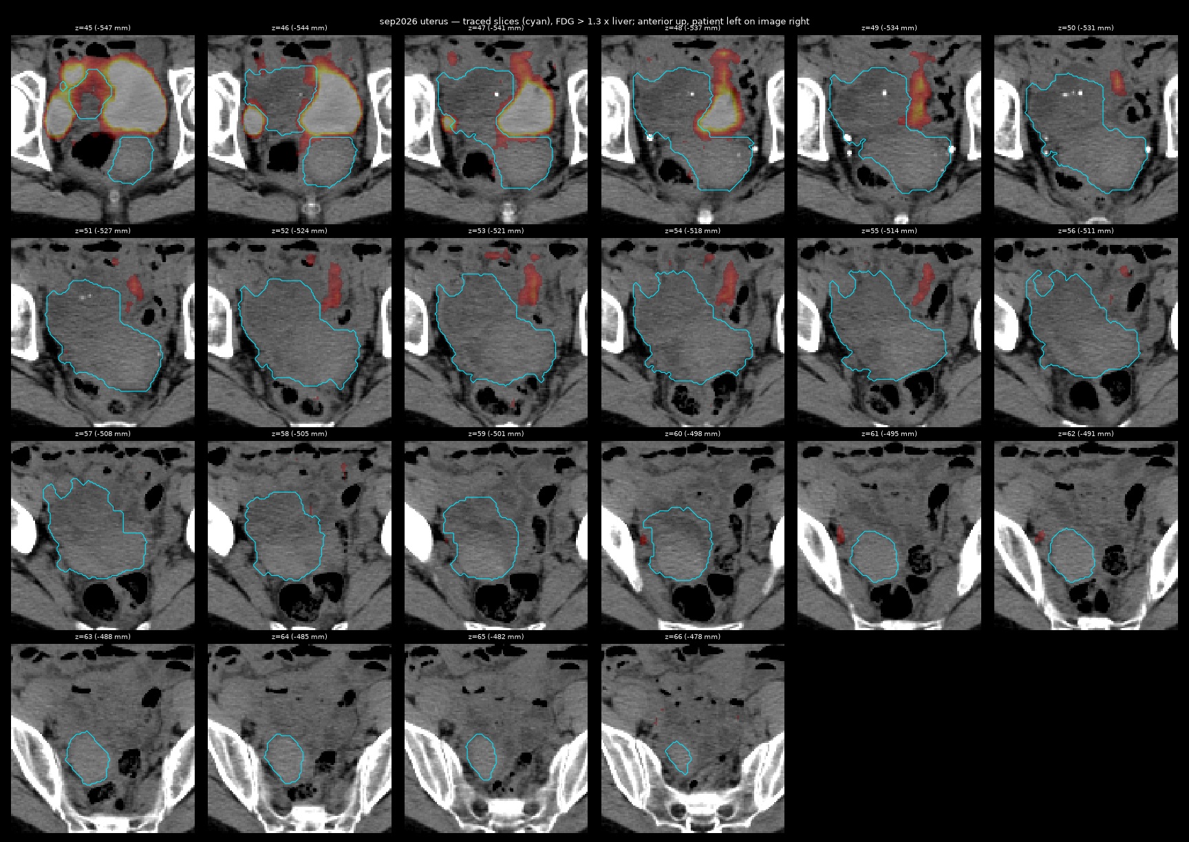
Femoral heads
| Site | Our measurement (June 2026 CT/PET) | Reports |
|---|---|---|
| Femoral heads | No lucent lesion found by our density criterion in either femoral head (see the bone-window montage) | LISOD 08.07.2026: new 31 mm right femoral-head lucency with sclerotic rim, moderate activity, not called metastasis; Israel 08.09.2026: no bone lesions; KMKOC PET-CT 23.09.2026: no bone lesions |
Femoral heads — bone window and FDG
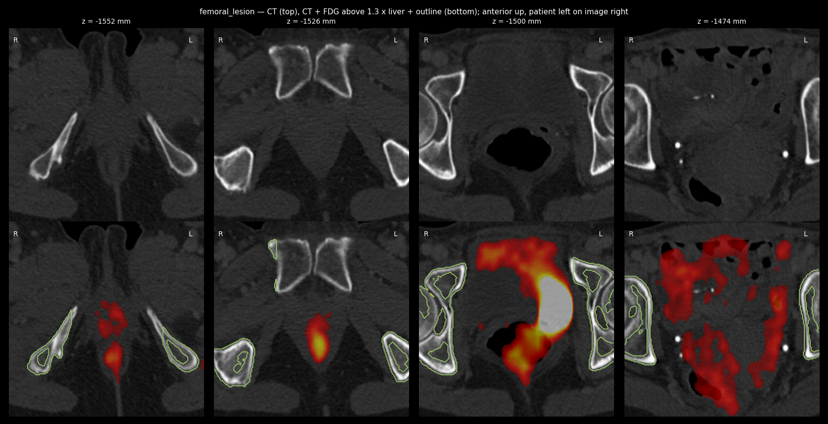
Static renders of the whole-body model
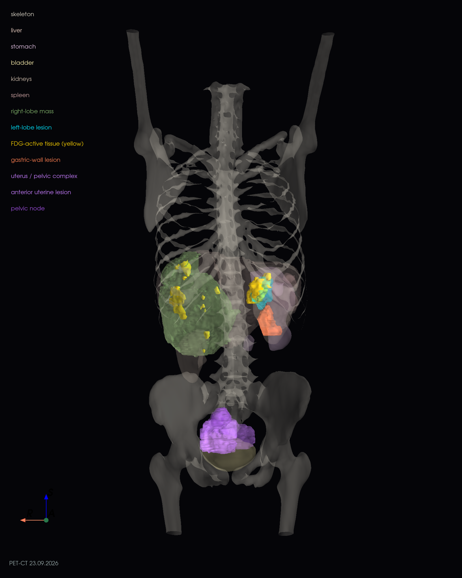
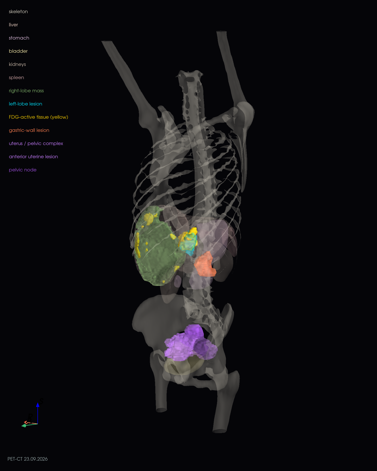
Interventional-radiology opinion — Dr. Oleksandr Korshak, head of the interventional radiology department, Shalimov National Scientific Centre of Surgery and Transplantology, Kyiv (profile), 10–11.09.2026, via messenger
Thermal ablation (RFA, MWA) is contraindicated for this lesion because two contraindications coincide: proximity to the left branch of the portal vein and, most importantly, direct adjacency to the left bile duct. Y-90 radioembolisation (TARE) costs from about EUR 40,000 and is not performed in Ukraine (nor in Czechia or Slovakia) for that reason. FACT Histotripsy at HCA Healthcare UK (London) costs from £37,500 for the treatment itself, plus £500 initial review (oncologist + MDT), about £250 per clinician per visit and about £100 for the radiologist's imaging review (e-mail reply of 11.09.2026; no quote for her case issued). The family's questions about transarterial therapies (DEB-TACE, TACE, TARE) and about surgical resection remain open.
Assessment against the imaging: consistent. The lesion abuts the left portal branch and the metabolically active tumour is exactly on that side, so an ablation margin would have to include the branch and the accompanying duct.
Histotripsy (11.09.2026, Dr. Korshak). Asked about focused-ultrasound histotripsy (HistoSonics Edison system, which has just received regulatory approval in Europe with a phased roll-out to selected centres — Focused Ultrasound Foundation, 2026; the company reports about 95 % local control after re-analysis, because tumour vessels persist and were previously mis-read as residual tumour), he answered that it is a new and very expensive method (HCA price list 11.09.2026, below) with no results yet in a large number of patients with this tumour type (GIST liver metastases); "liver tumours" is too broad a category, these tumours are too heterogeneous to transfer results from one tumour type to another, and there are no long-term results for tumours of this size either. He suggested asking HistoSonics directly whether a 4 cm GIST metastasis abutting the left portal pedicle is a candidate. Our follow-up question to him — whether the target would be the small new focus rather than the old tumour that imatinib still controls — is pending.
Where histotripsy can be asked for (September 2026). HistoSonics has no European office; enquiries go through the company's physician / general enquiry forms (patients via myhistotripsy.com); its VP Global Market Access, Joshua King, joshua.king@histosonics.com. Centres treating liver tumours outside trials: Addenbrooke's, Cambridge (Dr Teik Choon See, interventional radiology; NHS and private patients; cuh.histotripsyenquiries@nhs.net, private consultations cuh.private.addenbrookes@nhs.net, subject "Private Histotripsy"; +44 1223 348920) and The Wellington Hospital, London (HCA) (self-pay, quote per case; histotripsyenquiries@hcahealthcare.co.uk, +44 20 7908 8045). FACT HCA Healthcare UK reply, 11.09.2026 (e-mail from the Patient Liaison Officer, Specialist Service Line Hub, to the family's enquiry): histotripsy is a new service at HCA UK for liver cancer (HCC, cholangiocarcinoma), metastatic liver cancer (including colorectal metastases) and neuroendocrine tumours. They ask whether liver cancer (primary or secondary) is part of the diagnosis; age over 18; breast-feeding or pregnancy; general anaesthesia (the procedure requires it; has the patient been advised she cannot have it); access to scans and clinical letters for the initial review by an oncologist / MDT; ability to travel to London (the initial consultation may be virtual). Costs stated: initial review with oncologist and MDT £500.00; treatment itself starts at £37,500.00; pre- and post-operative consultant fees approx. £250.00 per clinician per visit; likely £100.00 charge for a radiologist to review the imaging for the MDT. No quote for her case was issued (no eligibility decision). Cost is high (see prices above). On the continent the HOPE4LIVER trial sites — Klinikum Braunschweig, Universitätsklinikum Magdeburg, Humanitas Milan, Vall d'Hebron Barcelona — have the system but had not announced commercial treatment as of September 2026; the CE mark of July 2026 allows a phased roll-out "beginning with select centres of excellence". No published histotripsy series includes GIST metastases.
Guideline and evidence basis for treating the focal progression locally
- NCCN Guidelines, Gastrointestinal Stromal Tumors (von Mehren M et al., NCCN Guidelines Insights: GIST, version 2.2022, J Natl Compr Canc Netw 2022;20(11):1204–1214, jnccn.org; NCCN Guidelines for Patients: GIST 2023, PDF): for limited progression on imatinib the options are to continue the TKI and add resection (if feasible), ablation, embolization or chemoembolization of the progressing lesion(s), with palliative radiotherapy for symptomatic lesions (category 2B); a switch to the next-line TKI is reserved for generalised progression. Resection of metastatic disease "may be beneficial in patients on imatinib or sunitinib who have evidence of radiographic response or limited disease progression".
- 2026-09-23 — PET-CT whole body (low-dose CT, no contrast) — English translation Imaging translation on Drive [hosted copy]
original (scanned PDF) - ESMO–EURACAN–GENTURIS clinical practice guideline (Casali PG et al., Ann Oncol 2022;33(1):20–33, doi:10.1016/j.annonc.2021.09.005): in case of limited progression on imatinib, treatment of the progressing lesion (surgery, ablation) with imatinib continued, or imatinib dose escalation, are the recommended options before changing the systemic drug.
- Dutch GIST Registry, multicentre analysis (Brink P, Kalisvaart GM, Schrage YM et al. Local treatment in metastatic GIST patients. Eur J Surg Oncol 2023;49(9):106942, doi:10.1016/j.ejso.2023.05.017, PubMed): of 457 metastatic GIST patients, 123 had elective surgery or ablation; local treatment was associated with better overall survival (HR 0.56, 95 % CI 0.34–0.93). After local treatment, disease confined to the liver predicted better survival (HR 0.27) and progression during systemic treatment predicted worse survival (HR 3.9) — so the benefit is largest when the disease is liver-limited and responding, and the current focal progression on imatinib is exactly the setting in which patient selection matters most.
- GIST Support International, "Radiofrequency ablation (RFA)" (gistsupport.org): RFA "is applicable to relatively small liver tumors that are not close to blood vessels or ducts in the liver" and can destroy a resistant clone while imatinib controls the rest; cited series: Dileo et al. 2004 (nine patients, limited progression sites, all lesions completely ablated, no complications), Hasegawa et al. 2007 (three patients with focal liver progression, progression-free at 8, 15 and 16 months), Pawlik et al. 2006 (RFA ± surgery vs surgery alone: higher recurrence with RFA, reflecting selection), Avritscher and Gupta 2009 (RFA "a viable palliative option for patients with advanced GIST who develop focal progression … during imatinib therapy"). The same source's proximity caveat is the reason thermal ablation was judged contraindicated here (contact with the left portal branch and bile duct), which leaves resection, SBRT, IRE or transarterial treatment as the guideline-compatible local options.
Evidence review (OpenEvidence conversation, 05.09.2026, 16 questions, 91 pages — PDF)
Bottom line: imatinib-refractory, liver-limited oligoprogression with polyclonal KIT resistance; local treatment of the active nodules with imatinib continued is favoured over a plain drug switch; fresh imaging (≤ 30 days old) is required before any procedure and the 32 % ctDNA fraction argues for acting within about a month.
All genomic findings from both tests
Full list of variants, fusions and genomic markers from the Oncomine (2022–2023) and FoundationOne (2026) reports
| Test | Finding | Allele fraction / level | Significance (as stated in the report) |
|---|---|---|---|
| Oncomine Comprehensive Assay Plus, CSD Kyiv, tissue block 48278/22 (Aug 2022 liver biopsy, ~80 % tumour cells), executed 06.01.2023 report 22OM011[11] · genetic commentary[18] · Mainz molecular tumour board[35] · raw BAM files (Drive) | KIT exon 11 deletion c.1668_1676delGTGGAAGGT (p.Gln556_Val559delinsHis = Q556_V559>H) | 42.5 % | Driver; sensitive to KIT inhibitors (imatinib, sunitinib, regorafenib, ponatinib named) |
| KIF5B–MET fusion (RNA) | 263 reads per million — subclonal | Potential MET-inhibitor sensitivity, judged unlikely to matter clinically; not a registered indication | |
| MTAT–CDKN2B-AS1 translocation; BRD3–NUTM1 translocation | — | No clinical or therapeutic significance | |
| TPMT p.Tyr240Cys; TPMT p.Ala154Thr | 39.8 % / 49.9 % | Pharmacogenetic (thiopurine metabolism); no therapeutic significance here | |
| PPM1D p.Gln360His | — | No clinical significance | |
| Tumour mutational burden | Low, 4.82 mutations/Mb | Low expected benefit from PD-1/PD-L1 inhibitors | |
| Microsatellite status | MSS | Low expected benefit from immunotherapy | |
| Loss of heterozygosity | Not determined | — | |
| Hereditary panel 22B104, CSD Kyiv, 15.12.2022 report[12] | Germline BRCA1, BRCA2, CHEK2, PALB2 (the four genes of the BRCA Plus panel) | — | No pathogenic or likely pathogenic variants in these genes |
| Tumour therefore judged potentially insensitive to PARP inhibitors (no HRR mutation found) | |||
| Result does not exclude variants outside the panel | |||
| FoundationOne Liquid CDx (blood, ctDNA), signed 20.08.2026, tumour type stomach GIST full report[75] · summary · full text ctDNA tumour fraction 32 % (high) — findings considered reliable | KIT exon 11 deletion Q556_V559>H | 17.0 % | Original driver |
| KIT exon 13 V654A | 8.8 % | Secondary resistance mutation (imatinib, avapritinib); ATP-binding pocket | |
| KIT exon 18 A829P | 4.0 % | Activation-loop resistance mutation; similar profile to V654A | |
| BRCA2 T3085fs*26 | 1.5 % | Germline testing recommended to distinguish somatic vs germline; relevant to PARP inhibitors, not directly to GIST | |
| CDKN2A rearrangement (intron 2) | 12.8 % | Tumour-suppressor loss; associated with more aggressive GIST | |
| MTOR A1459D | 0.21 % (low level) | Activating; possible mTOR-inhibitor sensitivity (low confidence at this level) | |
| Blood tumour mutational burden | 3 mutations/Mb (low) | — | |
| Microsatellite status | MSI-high not detected | — | |
Proposed systemic drugs — coverage of her three KIT clones
Merged from the FoundationOne Liquid CDx report[75] (therapy list, NCCN categories and resistance symbols, pages 1 and 9–14) and the OpenEvidence review (mutation-by-mutation activity, INTRIGUE and VOYAGER subgroup data, toxicity). Median PFS values are trial subgroup figures, not predictions for her. No single approved drug cleanly covers both resistant clones, which is why local treatment with imatinib continued and trial referral were rated the strongest strategies.
| Drug / approach | FoundationOne report | Exon 11 Q556_V559>H (driver, 17 %) | Exon 13 V654A (ATP pocket, 8.8 %) | Exon 18 A829P (activation loop, 4.0 %) | VEGFR / PRES-haemorrhage risk | Status and comment |
|---|---|---|---|---|---|---|
| Imatinib 400 mg/day (current) | Listed: extensive evidence of resistance (V654A, A829P) | Active | Resistant | Resistant | None (no VEGFR activity) | Continuing. Still controls the exon-11 bulk disease; guideline advice is to keep it through any local therapy (2-year PFS after ablation 75 % with TKI vs 29 % without). |
| Sunitinib 50 mg, 4 weeks on / 2 off | Listed: extensive evidence of resistance (A829P) | Active | Strong (median PFS 15.0 mo, INTRIGUE exon 11 + 13/14) | Resistant (median PFS 2.3 mo) | High: grade ≥ 3 hypertension 22.6 %; PRES boxed-level warning; one intracranial-haemorrhage death in INTRIGUE | Prescribed 30.07.2026 and 08.09.2026, not started; regorafenib chosen instead (dispensed 30.09.2026). Standard second line; covers only one of her two resistant clones; if used, reduced starting dose and strict home blood-pressure monitoring were advised. |
| Regorafenib 160 mg, days 1–21 of 28 | NCCN category 1 in GIST; no resistance symbol | Active | Preclinically weak, but clinically acceptable (median PFS 7.4 mo in ATP-pocket subgroup, VOYAGER) | Active (median PFS 6.7 mo) | High: grade ≥ 3 hypertension ~10–24 %; PRES in label; hepatotoxicity boxed warning; ~72 % dose modifications; hand-foot reaction | Named as an option on 08.09.2026; chosen — dispensed 30.09.2026 (Stivarga 40 mg × 84), not yet started. Best-validated third-line agent; the only approved single drug with some activity against both clones. |
| Ripretinib 150 mg/day | Listed: limited evidence of resistance (ATP-pocket variants) | Active | Reduced (median PFS 4.0 mo, exon 11 + 13/14) | Strong (median PFS 14.2 mo, exon 11 + 17/18) | Low: grade ≥ 3 hypertension 5.8 %; no PRES in label; weak VEGFR2 | Approved fourth line. In the INTRIGUE ctDNA subgroup with co-occurring ATP-pocket + activation-loop mutations (n = 22) it performed the same as sunitinib (HR 1.07). The lowest neurovascular risk of the active drugs. Price: US list price about USD 45,500 per 90 × 50 mg tablets, i.e. one month at 150 mg/day (Drugs.com price guide); European prices are negotiated nationally and not published; not registered in Ukraine (import only). |
| Sorafenib 400–800 mg/day | NCCN 2A "useful in certain circumstances"; no resistance symbol | Active | Active in vitro (reviews conflict; ATP-pocket sometimes classed resistant) | Active (class effect) | Moderate: lower hypertension rate than regorafenib; no hepatotoxicity boxed warning | Off-guideline-preferred; GIST evidence is retrospective (third/fourth line). Broadest in-vitro coverage of her three alterations. |
| Avapritinib | Listed: extensive evidence of resistance | Active | Resistant | Active | Cognitive effects, intracranial bleeding warning | Not suitable: did not beat regorafenib in VOYAGER and is resisted by V654A. |
| Nilotinib | NCCN 2A, other tumour type; listed as active against V654A and A829 | Active | No activity at clinical doses (2026 cell-line study) | No activity at clinical doses | Low VEGFR | Not recommended: negative phase III in GIST; "no biological rationale" for secondary-mutant GIST. |
| Ponatinib | NCCN 2A, other tumour type | Active | Resistant (V654A is its escape mutation) | Active | Arterial-occlusion and hypertension warnings | Poor fit for her mutations. |
| Dasatinib | No resistance symbol | Active | No mutation-specific data | No mutation-specific data | Low VEGFR | About 3-month PFS in later lines; no GIST mutation-specific efficacy data. |
| Velzatinib (IDRX-42) — selective KIT inhibitor, StrateGIST-1 (phase 1/1b; phase 3 StrateGIST 3 recruiting in Europe) | — (not in report) | Active (ORR 19 % in exon 11 primary; ctDNA VAF reduction > 99 % in 67 %) | Active (exon 13 ORR 17 %; ctDNA VAF reduction > 99 % in 80 %) | Active (exon 17/18 activation-loop ORR 23 %; VAF reduction in 83 %) | Selective KIT inhibitor (limited VEGFR activity); hypertension not a reported class signal so far | Trial only. ASCO 2026: second line median PFS 13.7 mo (ORR 40 %), third line 9.2 mo (27 %), fourth line and beyond 5.6 mo (24 %; 11.0 mo if no prior ripretinib/bezuclastinib). OS not mature. The one drug with reported activity against both of her resistance classes; eligibility and site access to be checked. |
| Bezuclastinib + sunitinib (Peak trial, phase 3) | — (not in report) | Active | Yes (sunitinib) | Yes (bezuclastinib) | Retains sunitinib VEGFR risk | Phase 3 Peak positive (PFS 16.5 vs 9.2 months, HR 0.50; ASCO 2026); FDA decision due 30.11.2026; not yet available outside the US programme. |
| Pimitespib (TAS-116, Jeselhy) — oral HSP90 inhibitor, a different drug class: degrades mutant KIT as an HSP90 client protein instead of blocking the kinase; 160 mg/day, 5 days on / 2 off (CHAPTER-GIST-301, Kurokawa et al., Ann Oncol 2022) | — (not in report) | Active regardless of mutation (PFS HR 0.51 overall) | Mutation-agnostic: exploratory PFS HR 0.52 in patients with KIT exon 13/14 or 17/18 secondary mutations | Same (exon 13/14 and 17/18 analysed together) | Low: no VEGFR activity, no hypertension or vascular signal reported in 119 pooled patients (pooled safety, Int J Clin Oncol 2025); typical toxicity diarrhoea 76 %, appetite loss 35 %, creatinine rise 34 %, nausea 31 %, night blindness 12 % (grade 1) | Approved in Japan only (June 2022, fourth line after imatinib, sunitinib and regorafenib); not registered in the EU, US or Ukraine. Phase 3 (n = 86): median PFS 2.8 vs 1.4 months with placebo; crossover-adjusted OS 13.8 vs 7.6 months (HR 0.42); no objective responses, benefit is disease stabilisation (disease-control rate 67 % in the Japanese expanded-access programme, Int J Clin Oncol 2025). Import via named-patient services: about €8,840 per pack of 40 × 40 mg (Everyone.org, September 2026) = 10 treatment days, i.e. roughly €17,700 per 28-day cycle. Assessment: the only approved agent whose activity does not depend on which KIT clone dominates and the lowest neurovascular risk, but the benefit is measured in weeks — a late-line or bridging option, not a substitute for local control. |
| Local therapy + continue imatinib (TACE / TARE / SBRT / IRE / resection) | — (not in report) | — | Ablates resistant clones directly | Ablates resistant clones directly | No new VEGFR exposure | NCCN-endorsed for limited progression; requires fresh imaging (≤ 30 days) and a tumour board; thermal ablation of the left-lobe lesion has been judged unsafe (see above), so the candidates are SBRT, IRE, DEB-TACE/TARE or resection of segments II–III. |
Full evidence summary (systemic options, local options, timing, immunotherapy)
- Pattern: imatinib-refractory, liver-limited oligoprogression ("nodule within a mass") driven by polyclonal secondary KIT resistance; the exon-11 bulk disease still responds to imatinib. A combined local-plus-systemic strategy is favoured over simply switching TKI, and imatinib should continue throughout any local therapy (stopping the TKI collapses the benefit of ablation).
- Systemic options: sunitinib covers V654A but not A829P; regorafenib and ripretinib cover A829P and, per VOYAGER ctDNA data, regorafenib remains clinically reasonable against V654A; sorafenib has the broadest in-vitro coverage but only retrospective GIST data. Prior PRES with intracranial haemorrhage raises the stakes for potent VEGFR inhibitors (sunitinib, regorafenib); if one is used, start at a reduced dose with tight blood-pressure monitoring. Trial referral is recommended because no approved single agent covers the full profile.
- Liver-directed options for a viable nodule inside a cystic mass: arterially directed therapies (DEB-TACE/TACE, Y-90 TARE; NCCN 2A) treat only perfused tissue; microwave ablation (after cyst aspiration) or cryoablation for a few well-defined nodules; SBRT for a perivascular/central nodule that is unsafe for a probe; IRE is the vessel/duct-sparing alternative for a nodule abutting major vessels or central bile ducts (GIST data anecdotal); RFA is the least suited technique in fluid-containing targets.
- Timing: ablation planning needs multiphase CT or MRI no older than about 30 days; the June scan is stale and the 32 % ctDNA fraction argues for re-imaging now and definitive local therapy within roughly a month, provided the disease is still oligoprogressive.
- Not realistic now: mRNA neoantigen vaccines and TIL therapy (no GIST data, low TMB); checkpoint inhibitors alone give about 16 % response in TKI-refractory GIST.
- Other: the 31 mm lucent right femoral-head lesion mentioned in the 08.07.2026 LISOD read should be characterised (MRI) because extrahepatic disease changes the value of arterial therapies; the 08.09.2026 expert reading found no bone lesions.
Surgical and local-treatment literature for the board (retrieved 14.10.2026; verified records)
Literature by topic and 199 numbered references
Literature retrieved live on 14.10.2026 (PubMed E-utilities, Europe PMC incl. preprints, ClinicalTrials.gov API, Crossref, label and guideline documents)
Method in brief: eleven topic searches plus one parallel search on GIST with malignant ascitic fluid; every record cited below was retrieved in this session (PubMed XML, Europe PMC JSON/full text, Crossref metadata, registry JSON, label pages or guideline PDF) and stored under literature_raw/; the quotes behind each number are in verification_tables.md; the per-query log with hit counts is LITERATURE_SEARCH_LOG.md. Preprints (bioRxiv/medRxiv/Research Square, Europe PMC source PPR) were searched for every topic: none relevant to this case was found, so no preprint is cited. Numbers below are abstract-level unless the full text was read (marked FT in the verification table). Nothing here is a recommendation.
Topic 1 — Limited (focal/oligo-) progression on imatinib treated locally with the TKI continued (19 queries; 40 abstracts opened)
| Record | Design, n | Key numbers (as retrieved) | Applicability to this case |
|---|---|---|---|
| Fairweather 2018 [14]; morbidity companion [44] | 2 centres, 400 operations / 323 patients on TKI | median PFS after surgery on imatinib: responsive 36, stable 30, unifocal progression 11, multifocal 6 months; OS not reached / 110 / 59 / 24 months; complications 27.5 %, major 17.5 % | sets the expected gain by response category; whether L1 + S8 focus + wall plaques is “unifocal” or “multifocal” is itself a board question |
| Cho 2019 [45] | propensity analysis, 90 patients with focal progression on imatinib 400 mg (38 resection + imatinib vs 52 escalation) | time to imatinib failure 24.2 vs 6.5 months; OS 53.2 vs 35.1 months; IPTW HR 0.36 / 0.58 | the only comparison against a systemic alternative; surgical group had lower burden |
| Mussi 2010 [46] | 80 patients | 2-year PFS 64.4 % (surgery at best response) vs 9.7 % (at focal progression); morbidity 16.3 % | the patient is in the focal-progression group |
| DeMatteo 2007 [47]; Raut 2006 [48] | 40 and 69 patients | focal resistance: median progression 12 months, 2-year OS 36 %; 12-month PFS 80 / 33 / 0 % for stable / limited / generalised progression | “focal” meant one growing lesion |
| Al-Batran 2007 [49] | 38 patients, 3 centres | focal vs generalised progression after salvage: median PFS 11.3 vs 2.5 months; secondary KIT mutations found only in progressing lesions (4 patients) | supports resecting the progressing clone |
| Hasegawa 2007 [50]; Tse 2012 [51] | 16 and 9 patients (resection/RFA for focal progression) | median TTP 5.5 months; complete resection better (P = 0.014); liver abscess and bile leak among complications | few, small lesions; gastric origin matches |
| Brink 2023, Dutch GIST Registry [36] | 457 metastatic patients, 123 locally treated | local treatment OS HR 0.56; after local treatment liver-confined disease HR 0.27, progression during systemic therapy HR 3.9 | this case carries both the favourable and the unfavourable feature |
| Bauer 2014 (EORTC-STBSG) [16]; Du 2014 RCT [52]; Park 2014 [53] | 239 metastasectomies; RCT closed at 41/210; 134 patients | median OS 8.7 y (R0/R1) vs 5.3 y (R2), debulking did not prolong survival; 2-year PFS 88.4 vs 57.7 % (P = 0.089); PFS 87.7 vs 42.8 months | responders, not progression |
| Wen 2024 [54]; Zhang 2025 [55]; Pearce 2026 [20]; Hompland 2024 [56] | 238 / 73 / 96 / 12 patients | 10-year OS 91.9 % with hepatectomy vs 61.1 % imatinib alone; multifocal progression HR 2.41; cytoreduction + systemic OS 10.8 vs 7.4 years; 42 % progression-free 3 years after stopping imatinib in oligometastatic CR | mixed indications; selection bias |
Topic 2 — The large fluid/necrotic mass: drainage, sclerosis, rupture, infection, “nodule within a mass” (28 queries; 45 abstracts opened; negative finding on drainage)
| Record | Design, n | Key numbers | Applicability |
|---|---|---|---|
| Shankar 2005 [17] | imaging review, 92 trial patients | new nodule within a mass in 21 of 39 recurrences; first sign of progression in 17 of 21 | the imaging pattern of this case; no management data |
| Choi 2007 [15] | 172 lesions / 40 patients | density/size criteria: sensitivity 97 % vs RECIST 52 % for PET response | why size-stable cystic masses count as response and new nodules as progression |
| Tirotta 2019 [4] | 470 TKI-treated patients | complicated tumour response (bleeding, abscess, perforation) in 6.4 %; percutaneous drainage in 4 of 30; mortality 13.3 %; median onset 14 months | only series quantifying spontaneous complications of responding disease |
| Colović 2013 [57]; Suzuki 2008 [58]; Radkani 2008 [59] | single cases, 14–20.5 cm cystic metastases | operative excision or partial excision then imatinib; PVE + lobectomy with post-operative subhepatic abscess | litre-scale analogues, all imatinib-naive, none drained percutaneously |
| Akbulut 2018 [60] | single case, giant cystic ileal GIST | repeated percutaneous drainage (≥ 10 L, then 6 L) → re-accumulation, infection, resection | the only published drainage course of a cystic GIST |
| Jang 2018 [61]; Denton 2026 [62]; Yu 2024 [63]; Pierce 2018 [64] | case reports / 28-patient embolisation series | rupture of hepatic GIST metastases spontaneously, after Y-90 and during a TACE course; arterial embolisation the usual rescue; ruptured GIST median OS 611 days | subcapsular lesions and interventions carry rupture risk |
| Abscess after TACE/ablation: Wang 2025 [65]; Song 2001 [66]; Ye 2022 [67]; Wang 2024 [68] | meta-analysis 254,408 TACE; 2,439 patients; 137 DEB-TACE for metastases; 314 DEB-TACE | pooled abscess 0.54 % (mortality 7.7 %); metastatic tumours 2.6 %; 8.8 % per patient in metastatic DEB-TACE; 3.8 % with large size as a risk factor | biliary intervention, the dominant risk factor, is absent here; metastatic histology and large size apply |
Topic 3 — Sequencing, staged hepatectomy, ALPPS/PVE, major hepatectomy for GIST metastases (25 queries; negative finding on lesion order)
| Record | Design, n | Key numbers | Applicability |
|---|---|---|---|
| Kraft 2023 [69] | single case, bilobar giant GIST metastases, TKI-refractory | two-stage debulking: left side first, right posterior sectionectomy seven weeks later; grade A liver insufficiency after stage 1; survival 55 months from diagnosis | the only published two-stage bilobar GIST hepatectomy; left-first order unexplained; no PVE, no R status |
| Liu 2017 [70] | single case, right-sided bulky necrotic metastases on imatinib | FLR 326 mL (34 %) → 50 % in 10 days after PVE plus microwave partition; right trisegmentectomy | remnant tumour-free, unlike L1 here |
| Lai 2021 [71] | systematic review of uncommon ALPPS indications, 136 cases | 6 GIST cases (4.4 %); Clavien–Dindo ≥ IIIa in 2 of 3 evaluable | ALPPS in GIST is anecdotal |
| Bowers 2012 [72]; Schadde 2014 [73]; Adam 2006 [74] | two-stage hepatectomy (39, GIST n = 1); ALPPS registry (202); 1,452 non-colorectal resections | morbidity 23 % and 56 % per stage, liver insufficiency 5 % and 22 %; ALPPS 90-day mortality 9 %, non-colorectal OR 2.7 for severe complications; R0 83 %, 60-day mortality 2.3 % | staged-surgery risk benchmarks from other tumours |
| Sutton 2022 [75]; Seesing 2016 [76]; Xue 2022 [77]; Shi 2017 [78]; Turley 2012 [79] | 91 / 48 / 119 / 144 / 39 patients | resection of treatment-resistant lesions: 10-year OS 58 % vs 72 % curative-intent vs 41 % unresected; major hepatectomy 25–39 %; R0 71–72 %; severe complications 10 %, mortality 2.5 %; extrahepatic disease HR 9.51 | responders with 4–5 cm lesions; no staged procedures |
| Zeng 2024 [80]; Chen 2019 [81]; Rahimi-Ardabily 2023 [21] | RFA vs hepatectomy cohorts; meta-analysis 988 patients | PFS 14.3 vs 42.7 months and 9 vs 29 months favouring resection; complications 10 % vs 53 % favouring RFA; resection + TKI OS HR 2.10 | modality trade-off in GIST liver metastases |
| Fan 2025 [82] | single case | resection of a 14 cm lesion carrying secondary KIT exon 13 V654A at limited progression on imatinib; no recurrence at 39 months | the only published resection of a V654A-bearing progressing lesion |
Topic 4 — Peritoneal disease and ascitic cytology: AUS, cell block, GIST cells in fluid, surgery with peritoneal disease (25 topic queries + 47 queries of the parallel ascites search)
| Record | Design, n | Key numbers | Applicability |
|---|---|---|---|
| Jamal 2024 [2]; Castilho da Silva 2024 [83]; Ahuja 2024 [84] | meta-analyses of the International System (TIS) | pooled risk of malignancy: AUS 49 % (95 % CI 37–61 %), malignant 100 %; AUS 50.8 %; specificity of “malignant” 99 %, of “atypical and above” 95 % | carcinoma-dominated data; the two readings here imply very different pretest risks |
| Doğan 2025 [27]; Wang 2023 [85] | 3,501 effusions; review | AUS ROM 27 %; inter-observer kappa for AUS 0.162; AUS is “a diagnosis of exclusion” needing clinical correlation; peritoneal-fluid AUS ROM about 22–63 % | disagreement between laboratories on an atypical fluid is documented and common |
| Yarikkaya 2025 [28]; Fatima 2011 [86] | 37 AUS fluids; 98 FNA cell blocks | cell-block immunopanel re-classified 86.1 % of AUS; DOG1 sensitivity 100 %, c-kit 70 % | the resolving test for GIST is a cell block with DOG1/CD117, not the usual epithelial panel |
| Sultan 2025 [3]; Tajima 2016 [30]; Wong 2002 [87]; Valdivia-Mazeira 2022 [88] | case reports (the entire direct evidence) | GIST cells in ascites/effusions were large, epithelioid or pleomorphic, mimicking adenocarcinoma; sparse in smears; diagnosis rescued by DOG1/CD117 on cell block | the cells described on review here were small and monomorphic |
| Shidham 2021 [29]; Chen 2019 [89]; Wieczorek 2001 [90] | reviews / series | false positives up to 0.5 %, false negatives up to 30 %; low-grade sarcomas rarely exfoliate; GIST aspirates pleomorphic in 7 % | a Ki-67 2 % GIST has a low tendency to shed cells; not GIST-specific evidence |
| Kim 2015 [26] | 403 imatinib-treated GIST | fluid retention in 15; ascites in 80 % of these; intermittent mild retention at any time | a benign explanation for small-volume ascites on long-term imatinib |
| Nishida 2019 [35]; Hølmebakk 2018/2019 [39,91] | rupture definition; Oslo cohorts | rupture criteria do not include positive cytology; incisional biopsy counts as rupture, needle biopsy does not; gastric GIST 5-year RFS 37 % after rupture; peritoneal penetration without rupture not prognostic | framework from localised disease; laparoscopic incisional biopsy of GF meets the definition |
| Li 2024 [19]; de la Fuente 2013 [92]; Pearce 2026 [20]; Rahimi-Ardabily 2023 [21] | 194 / 59 / 96 / 988 patients | peritoneum-only vs liver-only OS HR 2.67; liver-only vs liver + sarcomatosis OS 40.5 vs 28.7 months (NS); no OS difference by site in oligometastatic disease; hepatectomy benefit retained in studies with concurrent peritoneal disease | peritoneal disease worsens prognosis but did not abolish the association of liver surgery with survival |
| Gutiérrez 2026 [31]; Spiliotis 2026 [32]; Bryan 2014 [93]; López-Rojo 2026 [94] | peritoneal GIST surgery series | 5-year OS 77.8 %; median OS 62 months, better with PCI ≤ 10; TKI + cytoreduction 7.9 years vs 1.04 without TKI; progressing on TKI before surgery 1.35 years | macroscopic peritoneal disease; cytology never a variable |
| Hariharan 2012 [95] | meta-analysis, colorectal liver metastases | staging laparoscopy yield 19 %, sensitivity 75 % for peritoneal lesions | no GIST-specific data on staging laparoscopy |
Topic 5 — Ablation next to the portal pedicle and bile ducts: IRE, MWA/RFA, histotripsy; GIST ablation series (37 queries; 72 abstracts; 3 full texts; histotripsy regulatory documents)
| Record | Design, n | Key numbers | Applicability |
|---|---|---|---|
| Silk 2014 [5] | IRE within 1 cm of major hepatic ducts, 11 patients / 22 metastases (1.0–4.7 cm) | transient liver-test rise after 4 sessions; 1 stent; prolonged abnormalities attributed to progression | closest anatomical match; no efficacy data; not GIST |
| Thamtorawat 2022 [6] | IRE 13 vs RFA 31 peribiliary tumours (~1.8 cm) | significant biliary complications 7.7 % vs 16.1 %; local progression 23 % vs 44 % at 3 years (NS) | direct IRE-vs-thermal comparison at the duct; lesions far smaller than 5 cm |
| Dollinger 2016 [96]; Distelmaier 2017 [97]; Tamura 2020 [98] | 55 ducts within 1 cm of IRE zones; 43 tumours against major veins; vessel occlusion study | radiological duct changes common but ducts “largely unaffected”; complete ablation 93 %, cholestasis in 24 % of portal-adjacent cases, tract recurrence 26 %; vessels ≥ 4 mm rarely occlude | a 5 cm zone would encase segmental branches |
| COLDFIRE-2 [99]; IRE pooled data [100,101,102] | prospective CRLM ≤ 5 cm (n = 50); 807 / 776 / 180 patients | 1-year local-progression-free 68 %, adverse events 40 %, 1 death; complete ablation 86 %, complications 23 % (major 6.9 %); biliary stricture 2.8 % | the only prospective IRE data admitting 5 cm lesions; colorectal |
| van Tilborg 2016 [103]; Felker 2015 [104]; Ohnishi 2008 [105] | MWA vs RFA peribiliary CRLM (199 lesions); biliary cooling series | biliary complications “especially common” after peribiliary MWA (P = 0.002); cooling reduced biliary injury from 31 % to 0 % and from 46 % to 2.5 % | thermal ablation at a first-order duct is not an absolute contraindication in cooled, small lesions |
| GIST RFA series: Jung 2015 [106]; Hakimé 2014 [107]; Yamanaka 2013 [108]; Yoon 2018 [109]; Liu 2024 [110]; Yang 2020 [111] | 29 / 17 / 7 / 24 / 34 / 55 patients | technical effectiveness 95.6 % at mean 1.3 cm (1 tract seeding); 2-year PFS 75 % with TKI maintained vs 29 % discontinued; local progression 4.8 %; biliary stricture and abscess among major complications; ablation vs resection OS 102 vs 117 months, complications 18.5 % vs 78.6 % | same clinical setting; lesions a quarter of L1’s size, no duct contact; peri-organ lesions left untreated |
| Histotripsy: #HOPE4LIVER [8,42]; multicentre data [112,113,114]; near-vessel data [43]; sarcoma case [115]; regulatory [116,117]; trials [118,119,120] | pivotal 44 patients / 49 tumours ≤ 3 cm; 230–972 patients; 21 tumours abutting major veins; 1 liposarcoma case | technical success 95 %; 1-year local control 63.4 % (90 % post hoc); major complications 1.3–7 %; 30-day mortality 11.1 % in a US practice cohort; bland portal vein thrombosis 38 % when zones abut large veins; CE Mark 13 July 2026 with no labelled size limit; FDA order states the device “has not been evaluated for the treatment of any specific disease” | no GIST treated; hilar structures excluded from pivotal treatment volumes; no European liver histotripsy trial recruiting |
Topic 6 — TARE/SIRT, TACE, DEB-TACE and bland embolisation in GIST (21 queries; 5 full texts)
| Record | Design, n | Key numbers | Applicability |
|---|---|---|---|
| Hohenberger 2025 [24]; Rathmann 2015 [121]; conference abstract [122] | SIRT, 26 TKI-resistant patients (median 3 prior lines); precursor 9–11 | Choi response 16/26; median hepatic PFS 16 months, OS 28 months; KIT exon 11 with secondary mutations: hepatic PFS 7 months; 3 gastric ulcers needing surgery; 2 excluded for lung shunt > 20 % | TKI-exhausted population; the index genotype had the shortest hepatic PFS; no lesion-size data |
| Berman 2026 [123] | SIRT, 12 SDH-deficient GIST | ORR 66.7 %, DCR 100 % | different biology |
| Liu 2026 [25]; Cao 2012 [124]; Cao 2014 [125] | DEB-TACE 34 vs 43 (≥ 2 TKI lines); cTACE 22 vs 38; bland vs cTACE 45 | ORR 94 %, liver PFS 11.0 vs 5.0 months, OS 18 vs 12 months; liver PR 54.5 %, OS 68.5 vs 25.7 weeks; PFS 56.6 vs 42.1 weeks | all after TKI failure; lesions selected or described as hypervascular; cystic lesions showed only faint ring staining |
| Kobayashi 2006/2009 [126,127]; Takaki 2014 [128]; Maluccio 2006 [129] | 110 HACE; 14 imatinib-resistant; 11 bland HAE; 24 sarcoma (16 GIST) | PR 14 %, liver PFS 8.2 months, OS 17.2 months; imatinib-resistant liver PFS 7.0 months; bland HAE PFS 3.9 months, no major complications; response 60 % | largely pre-imatinib or late-line |
| Kamat 2008 [130]; Miller 2018 [131]; Pierce 2018 [64] | 60 patients with > 75 % liver involvement; 39 hepatic sarcoma Y-90; 28 sarcoma embolisations | major complications 19 %, 12 procedure-related deaths with massive burden; Y-90 response 36 %, OS 30 months; ruptured GIST OS 611 days | the only series addressing very large tumour burden warns against embolisation with massive burden plus risk factors |
| Malavia 2026 [132]; Yerubandi 2016 [133]; Denton 2026 [62]; Wang 2024 [68] | hypovascular Y-90 (27); shunt predictors (190); rupture case; abscess 314 | hypovascular tumours still showed tumour-to-normal uptake ratios > 1 (necrotic tumours excluded); shunt > 10 % predicted by tumour diameter; rupture one day after SIRT; abscess 3.8 % with large size as risk factor | addresses the “dark on CT” objection only indirectly |
Topic 7 — Imatinib around surgery; regorafenib/sunitinib/ripretinib holds; VEGFR TKIs after PRES (33 queries; labels and guideline texts read)
| Record | Design, n | Key statements and numbers | Applicability |
|---|---|---|---|
| ESMO–EURACAN–GENTURIS 2022 [11]; GEIS 2023 [38] | guidelines (full text read) | surgery or ablation of focal progression (“nodule within a mass”) with imatinib continued [IV, C]; imatinib “can be safely stopped a few days or even 1 day before surgery, to be resumed promptly”; GEIS: stop 24 h before, restart once oral tolerance is confirmed; interruption “generally followed by relatively rapid tumour progression” [I, A] | direct answers to Q6 on imatinib timing |
| BFR14: Blay 2007 [134]; Le Cesne 2010 [13]; Patrikidou 2013 [135]; Blay 2024 [136] | randomised interruption after 1, 3 and 5 years | 2-year PFS 80 % vs 16 % after 3-year interruption; median PFS 6.1 vs 27.8, 7.0 vs 67.0, 12.0 months vs not reached; only 42 % of CR patients regained CR on rechallenge; interruption “not recommended” | planned months-long stops, not peri-operative pauses |
| Metastasectomy on TKI: Fairweather 2018 [44]; Raut 2006 [48]; Mussi 2010 [46]; Hasegawa 2007 [50]; Raut 2010 (sunitinib) [137]; Lee 2019 [10]; Rajkumar 2006 [138] | 400 operations; 69; 80; 16; 50 on sunitinib; 613 (108 pre-operative TKI); mouse model | complications 27.5 %, major 17.5 %; morbidity 16.3 %; no hospital deaths; surgery on sunitinib: complications 54 %, reoperation 16 %; pre-operative TKI: complications 34 % vs 10 %, anastomotic leak 18 % vs 6 %; imatinib delayed wound healing in mice at 7 days, closed by 14 | no series states the day imatinib was stopped; the imatinib label has no hold statement |
| Labels: regorafenib [12,139]; sunitinib [140,141]; ripretinib [142,143] | current US and EU product information | regorafenib: withhold ≥ 2 weeks before elective surgery, ≥ 2 weeks after major surgery until healing; RPLS → discontinue; BP must be controlled before starting; sunitinib: ≥ 3 weeks before, ≥ 2 weeks after; RPLS → discontinue (US) / temporary suspension (EU); ripretinib: ≥ 1 week before (US) or 5 days before and after major surgery (EU); no PRES statement | the holds define the earliest regorafenib start relative to each operation |
| PRES rechallenge: Tlemsani 2016 [144]; Miller-Patterson 2017 [145]; Lee 2026 [146]; Imai 2022 [147]; Catalano 2022 [148]; Deguchi 2018 [149]; Li 2026 [150] | 34 neurovascular events; case reports; FAERS | severe vascular toxicity recurred in 2 of 6 reintroductions; pazopanib PRES with occipital haemorrhage: no anti-VEGF drug restarted; pazopanib four weeks after a sunitinib-related cerebellar bleed: fatal re-bleed; lenvatinib after sorafenib RPLS: no recurrence over 5 years; PRES recurred on sunitinib reintroduction; pazopanib restarted at half dose under BP control without recurrence; PRES signal for 13 of 16 VEGF-pathway agents | no report of any VEGFR TKI after PRES with intracerebral haemorrhage; none of regorafenib or ripretinib after PRES |
| Blood pressure: Maitland 2010 [151]; ESC 2022 [152]; RT + VEGFR TKI toxicity: Brade 2016 [153]; RT with TKI continued: Joensuu 2015 [7]; SBRT case [154] | consensus; guideline; phase I; phase II; case | goal < 140/90 mmHg (< 130/80 with diabetes); home monitoring daily in the first cycle; hold at ≥ 180/110; sorafenib with SBRT “not recommended outside a clinical trial”; conventional RT with TKI unchanged well tolerated; SBRT 42 Gy/6 fractions with sunitinib continued | frames Q7; no post-PRES-specific target exists |
Topic 8 — KIT exon 13 V654A and exon 18 A829P, inter-lesion heterogeneity, clone-targeted local therapy (23 queries; 31 abstracts; 3 full texts)
| Record | Design, n | Key numbers | Applicability |
|---|---|---|---|
| Heinrich 2024, INTRIGUE ctDNA [1] | randomised second-line trial, ctDNA in 362 | exon 11 + 13/14: sunitinib PFS 15.0 vs ripretinib 4.0 months; exon 11 + 17/18: ripretinib 14.2 vs 1.5 months; co-occurring pocket + loop mutations (n = 22): HR 1.07, no difference; V654A the commonest secondary mutation | the index profile is the subgroup without a preferred drug |
| Serrano 2023, VOYAGER ctDNA [155] | 386 ctDNA samples | ATP-pocket mutations: avapritinib PFS 1.9 vs regorafenib 5.6 months; regorafenib unaffected; 23.4 % carried both pocket and loop mutations | argues against avapritinib; regorafenib indifferent to a pocket clone |
| Serrano 2019 [22] | cell lines and xenograft | V654A: sunitinib 45 nM vs regorafenib 2000 nM; A829P: no agent below the authors’ 500 nM threshold (regorafenib 2500 nM); ripretinib and avapritinib not tested | the only comparative sensitivity data for both mutations; no clinical A829P series |
| Liegl 2008 [33]; Wardelmann 2006 [156]; Li 2025 [40]; Mühlenberg 2024 [157] | 53 metastases / 14 patients; 104 samples / 32; 206 samples / 56; 25 ripretinib-refractory | 67 % had different secondary mutations in different metastases; 43.8 % acquired new mutations; a secondary mutation was present in only 57.5 % of samples from mutation-positive patients, and in 95 % vs 32 % depending on continued imatinib; pocket + loop mutations often in cis | two plasma mutations do not say whether L1 carries one, both or neither |
| Desai 2007 [158]; Al-Batran 2007 [49]; Cho 2019 [45]; Ono 2019 [159]; Fan 2025 [82] | imaging series; 38; 90; cases | resistant nodule precedes overall progression by 5 months and carried a new mutation in 8 of 10 biopsies; secondary mutations only in progressing lesions; resection at focal progression TTF 24.2 vs 6.5 months; resected focal nodule carried exon 18; resected V654A lesion without recurrence at 39 months | rationale and case-level precedent for removing the resistant clone |
| Johansson 2021 [41]; Hashimoto 2024 [160]; Heinrich 2008 [161] | 161 plasma samples / 32; 133; 97 on sunitinib | ctDNA cleared after surgery in all positives; ctDNA-matched TKI PFS 8.23 vs 2.43 months; exon 13/14 better than 17/18 on sunitinib | ctDNA is a plausible monitoring tool after local therapy; no validated endpoint |
Topic 9 — The gastric primary after TKI: pathological response, calcification, resection of the primary, combined procedures (32 queries)
| Record | Design, n | Key numbers | Applicability |
|---|---|---|---|
| Bauer 2005 [9]; Rutkowski 2006 [162]; Scaife 2003 [163]; Agaram 2007 [18]; Vassos 2021 [164]; Goh 2006 [165] | resected responders (11, 24, 17 specimens); 43 nodules; 50 neoadjuvant gastric; review | viable cells in all but one, 21 of 24 and 15 of 17 specimens; histological response heterogeneous and unrelated to duration; pCR 24 % after neoadjuvant imatinib; pathological response does not correlate with PET | one hyalinised 1.5 cm fragment cannot exclude viable foci in a 4 cm lesion |
| Hedenström 2017 [166]; Kondo 2018 [167]; Cameron 2010 [168] | biopsy vs specimen Ki-67; 6-year pCR case; mirror-image case | Ki-67 2.5 % on EUS vs 0.2 % on specimen; whole-specimen pCR after 6 years; primary regressed while liver metastases progressed with secondary mutations | no report of a calcified FDG-negative primary regrowing on continued imatinib |
| Warakaulle 2006 [169]; Werewka-Maczuga 2017 [170]; Eslamy 2008 [171] | imaging series / case | calcification occurs in responding lesions and increases on imatinib; one case of calcification increasing together with FDG activity | calcification is a response feature, not proof of cell kill |
| Zhao 2020 [172]; Gaitanidis 2019 [173]; Sato 2016 [174]; An 2013 [37] | SEER 455; propensity 570; registry 14; 249 | primary-site surgery OS 72 vs 40 months (HR 0.58), gastric subgroup HR 2.95 for no surgery; upfront primary surgery or cytoreduction before imatinib without benefit | registry data lack response and timing; no study resects a quiescent primary while metastases remain viable |
| Lee 2019 [10]; Fairweather 2018 [44] | 613 (108 pre-operative TKI); 400 metastasectomies | complications 34 % vs 10 %, anastomotic leak 18 % vs 6 % with pre-operative TKI; major complications 17.5 %, rising with complexity | supports the surgeon’s reluctance to combine a gastric wedge with a major hepatectomy |
| ESMO 2022 [11] | guideline | surgery “not recommended as a primary approach in the metastatic setting”; residual-disease surgery “should be individualised” [III, C] | — |
Topic 10 — Tumour spillage and seeding after puncture, biopsy or drainage of GIST (33 queries; ESMO text read; negative finding on drainage)
| Record | Design, n | Key numbers | Applicability |
|---|---|---|---|
| Jakob 2022 [175]; van Houdt 2021 [176]; Eriksson 2016 [177] | systematic review 350 patients; cohort 228; trial subset 47 biopsied | one needle-tract seeding after percutaneous core biopsy; 1 of 70 transcutaneous biopsies (1.4 %); route not significant in multivariable analysis; no excess recurrence after biopsy under adjuvant imatinib | primary tumours before surgery; no liver-metastasis or cystic-lesion data |
| ESMO 2022 [11]; Nishida 2019 [35]; Hølmebakk 2016/2018 [39,178]; Joensuu 2024 [179] | guideline; definitions; cohorts | peritoneal contamination from a properly performed core biopsy “negligible”; cystic masses to be biopsied only at specialised centres; incisional biopsy counts as rupture, uncomplicated needle biopsy does not; 5-year RFS 37 % after rupture; 10-year RFS 21 % vs 55 % | rupture framework from non-metastatic disease |
| Davies 2008 [180]; Shoup 2002 [181] | case; 1,650 laparoscopies | port-site metastasis about 22 months after diagnostic laparoscopy with biopsy of peritoneal seedlings in gastric GIST on imatinib; port-site implantation 0.79 % after diagnostic laparoscopy for upper-GI malignancy | the closest analogue to the 05.10.2026 laparoscopy |
| Minoda 2023 [182]; Gao 2020 [183]; Terasawa 2023 [184]; Silva 2008 [185]; Berger-Richardson 2017 [186]; Iemoto 1981 [187] | EUS sampling; reviews; pancreatic EUS seeding; HCC meta-analysis; retroperitoneal sarcoma; case | tumour cells in gastric juice after incisional biopsy 64 % vs 0 % after EUS-FNA; no GIST EUS seeding case; cystic/necrotic components raised seeding to 3.7 % vs 0 %; HCC tract seeding 2.7 %; sarcoma 0.37 %; carcinomatosis after cyst aspiration | context from other histologies; no report of seeding after aspiration or drainage of a cystic GIST metastasis |
Ongoing trials relevant to this case (ClinicalTrials.gov API records opened 14.10.2026; eligibility must be confirmed by the sites)
| Trial | Status (14.10.2026) | Design | Sites of interest | Eligibility notes relevant here |
|---|---|---|---|---|
| StrateGIST 3, NCT07218926 [23] | recruiting | phase 3, velzatinib (IDRX-42) vs sunitinib after imatinib | Hungary (Budapest, Pécs), Germany (Berlin, Essen, Mannheim, Munich, Cologne, Dresden, Tübingen), UK; Poland (Kielce, Poznań, Warsaw) listed as recruiting in the registry record of 05.10.2026, but a parallel CTIS check (lit_trials_centres) reports Polish recruitment ended 12.10.2026 — confirm with the sponsor | progression on or intolerance to first-line imatinib; tissue KIT/PDGFRA result; no posted haemorrhage/PRES exclusion; PET-only progression and planned liver surgery need sponsor adjudication |
| Inlexisertib + ripretinib, NCT05957367 [188] | recruiting | phase 1/2, second line after imatinib | Frankfurt, Hamburg | measurable disease; uncontrolled hypertension excluded |
| Velzatinib expanded access, NCT07825701 [189] | available | individual patient access after ≥ 3 lines | GSK portal | ascites counts as unstable liver disease |
| Peak, NCT05208047 [190]; INSIGHT, NCT05734105 [191] | European sites closed / active not recruiting | bezuclastinib + sunitinib; ripretinib vs sunitinib | Poland, Czechia, Germany, UK (closed) | INSIGHT excludes ctDNA exon 13 (her V654A) |
| PATHFINDERS, NCT07109024 [192] | not yet recruiting | PET-guided lesion-level resistance profiling | Essen, Lübeck | fourth line only |
| OligoRARE, NCT04498767 [193] | recruiting (record 2024) | SBRT for rare oligometastatic cancers (sarcoma listed) | Warsaw, Gdańsk, Hamburg, UK | no carcinomatosis or effusion; lesions ≤ 6 cm — likely excludes R1 |
| Histotripsy: #HOPE4LIVER EU/UK NCT04573881 [118], US NCT04572633 [119], BOOMBOX NCT06486454 [120] | closed / closed / US registry recruiting | device trials and registry | Germany, UK, Italy, Spain (closed); US | ≤ 3 tumours of ≤ 3 cm; portal vein/bile ducts excluded from the treatment volume; no European liver histotripsy trial recruiting |
| VALOROUS, NCT06103669 [194]; RT + ripretinib, NCT06507683 [195]; NB003, NCT07379047 [196]; ctDNA-guided sunitinib/regorafenib, NCT05366816 [197]; NN3201 ADC, NCT06805825 [198]; ATEZOGIST, NCT05152472 [199] | recruiting (US / China / China–Korea / US / US / France) | local ablation or SABR of oligoprogressive lesions with systemic therapy continued; radiotherapy + ripretinib (fourth line); NB003 after imatinib (intracranial haemorrhage within 1 year excluded); exon 13 → sunitinib, exon 17 → regorafenib; KIT ADC; imatinib ± atezolizumab (fourth line) | outside reachable geography or later lines |
Most similar published cases and series (all topics)
| Citation | Similarity | What matches | What differs |
|---|---|---|---|
| Cho 2019 [45] | high | focal progression on imatinib 400 mg; resection with imatinib continued vs escalation | not liver-specific; no genotype |
| Fairweather 2018, unifocal-progression group [14] | high | surgery during unifocal progression on imatinib: PFS 11, OS 59 months | multi-organ; 2001–2014 |
| Heinrich 2024, co-occurring subgroup [1] | high | ctDNA with pocket + loop mutations on exon 11 background (n = 22): no preferred drug | drug comparison, not local therapy |
| Kraft 2023 [69] | medium | bilobar giant GIST metastases, two-stage resection, left side first, 7-week interval | TKI-refractory man, debulking, no PVE |
| Liu 2017 [70] | medium | right-sided bulky necrotic metastases on imatinib; PVE + partition; remnant 34 → 50 % | remnant tumour-free |
| Fan 2025 [82] | medium | resection of a V654A-bearing progressing lesion at limited progression | solitary 14 cm lesion |
| Colović 2013 [57] | medium | woman, gastric GIST, 20.5 cm cystic right-lobe metastasis | imatinib-naive; partial excision only |
| Davies 2008 [180] | medium | gastric GIST with peritoneal seedlings biopsied at diagnostic laparoscopy; port-site metastasis later | treatment-naive at laparoscopy |
| Sultan 2025 / Tajima 2016 / Wong 2002 [3,30,87] | medium | GIST cells in ascites identified by DOG1/CD117 on cell block | gross disseminated disease; pleomorphic cells |
| Miller-Patterson 2017 [145] | high (index event) | pazopanib PRES with occipital haemorrhage, resolved | renal cancer; no later anti-VEGF drug |
| Lee 2026 [146] / Imai 2022 [147] | medium | second VEGFR TKI after TKI-associated brain haemorrhage (fatal) / after non-haemorrhagic RPLS (no recurrence) | different drugs and tumours |
| Silk 2014 [5]; Thamtorawat 2022 [6] | medium | ablation within 1 cm / 5 mm of major ducts (IRE) | not GIST; smaller lesions |
| Hohenberger 2025, exon 11 + secondary subgroup [24] | medium | SIRT for TKI-resistant liver metastases; hepatic PFS 7 months in the index genotype | TKI-exhausted; no size data |
| Reddy 2025 [154] | medium | woman in her 40s, solitary oligoprogressive liver lesion, SBRT with TKI continued | sunitinib; one lesion |
Search-log summary (PRISMA-style counts) and verification
See LITERATURE_SEARCH_LOG.md for every query (database, exact string, date, hits, screened, included) and verification_tables.md for the per-citation retrieval status, DOI resolution, full-text status, retraction/correction flags and the quote behind each number. Summary counts are generated from those files and inserted at build time below.
PRISMA-style summary (queries run 2026-10-14; definitions in LITERATURE_SEARCH_LOG.md):
| Topic | queries | hits (sum, overlapping) | titles screened | abstracts/records assessed | included in citation list |
|---|---|---|---|---|---|
| T1 | 19 | 1339 | 205 | 40 abstracts | 23 |
| T2 | 28 | 3156 | 278 | 45 abstracts, 3 full pages | 21 |
| T3 | 25 | 672 | 337 | about 40 abstracts, 5 full texts | 21 |
| T4 | 26 | 1104 | 396 | about 45 abstracts, 2 full texts | 28 |
| T5 | 37 | 2434 | 458 | 72 abstracts, 3 full texts, 7 registry records | 35 |
| T6 | 21 | 2591 | 219 | about 35 abstracts, 5 full texts | 19 |
| T7 | 33 | 2690 | 413 | about 60 abstracts, 4 full texts, 4 labels, 2 guidelines | 27 |
| T8 | 23 | 621 | 266 | 31 abstracts, 3 full texts | 15 |
| T9 | 32 | 1677 | 241 | 52 abstracts, guideline PDF | 22 |
| T10 | 33 | 4170 | 388 | about 45 abstracts, 3 full texts, guideline PDF | 17 |
| T11 | 31 | 307 | 307 | 32 registry records | 16 |
| A | 47 | 1133 | see file | 42 abstracts, 6 full texts (worker’s statement) | 45 records in SOURCES.md; 30 re-retrieved here |
| Total | 355 | 21894 | 3508 | — | 208 citations (all kinds; a citation may serve several topics) |
Preprint servers (bioRxiv/medRxiv/Research Square via Europe PMC SRC:PPR) were queried for every topic; no relevant preprint was found, so none is cited. Conference abstracts (ASCO/ESMO/CTOS/CIRSE/SIR 2024–2026) were searched by web search only; none relevant was found except one 2014 RSNA abstract, cited as such.
References (numbered by first citation; Vancouver style; entries generated from the retrieved records)
- Heinrich MC, Jones RL, George S, Gelderblom H, Schöffski P, von Mehren M, et al. Ripretinib versus sunitinib in gastrointestinal stromal tumor: ctDNA biomarker analysis of the phase 3 INTRIGUE trial. Nat Med. 2024;30(2):498-506. doi:10.1038/s41591-023-02734-5. PMID 38182785. — Full text (open access, PMC)
- Jamal FE, Vey JA, Proctor T, Ishak A, Schmitt FC, Nikas IP. The International System for Reporting Serous Fluid Cytopathology: A Systematic Review and Meta-Analysis of Diagnostic Test Accuracy Studies. Adv Anat Pathol. 2024;31(4):256-264. doi:10.1097/PAP.0000000000000454. PMID 38695284. — Full text (subscription)
- Sultan H, Sadeghian Z, Goez-Gutierrez E, Diacovo MJ, Yoon E. Cytological Assessment of Metastatic Gastrointestinal Stromal Tumor in Ascitic Fluid. Diagn Cytopathol. 2025;53(9):462-465. doi:10.1002/dc.25499. PMID 40556369. — Full text (open access, PMC)
- Tirotta F, Fumagalli E, Colombo C, Morosi C, Barisella M, Radaelli S, et al. Management of complicated tumor response to tyrosine-kinase inhibitors in gastrointestinal stromal tumors. J Surg Oncol. 2019;120(2):256-261. doi:10.1002/jso.25491. PMID 31066052. — Full text (publisher, OA)
- Silk MT, Wimmer T, Lee KS, Srimathveeravalli G, Brown KT, Kingham PT, et al. Percutaneous ablation of peribiliary tumors with irreversible electroporation. J Vasc Interv Radiol. 2014;25(1):112-8. doi:10.1016/j.jvir.2013.10.012. PMID 24262034. — Full text (subscription)
- Thamtorawat S, Patanawanitkul R, Rojwatcharapibarn S, Chaiyasoot W, Tongdee T, Yodying J, et al. Biliary complications and efficacy after ablation of peribiliary tumors using irreversible electroporation (IRE) or radiofrequency ablation (RFA). Int J Hyperthermia. 2022;39(1):751-757. doi:10.1080/02656736.2022.2079733. PMID 35649727. — Full text (publisher, OA)
- Joensuu H, Eriksson M, Collan J, Balk MH, Leyvraz S, Montemurro M. Radiotherapy for GIST progressing during or after tyrosine kinase inhibitor therapy: A prospective study. Radiother Oncol. 2015;116(2):233-8. doi:10.1016/j.radonc.2015.07.025. PMID 26228971. — Full text (publisher, OA)
- Mendiratta-Lala M, Wiggermann P, Pech M, Serres-Créixams X, White SB, Davis C, et al. The #HOPE4LIVER Single-Arm Pivotal Trial for Histotripsy of Primary and Metastatic Liver Tumors. Radiology. 2024;312(3):e233051. doi:10.1148/radiol.233051. PMID 39225612. — Full text (open access, PMC)
- Bauer S, Hartmann JT, de Wit M, Lang H, Grabellus F, Antoch G, et al. Resection of residual disease in patients with metastatic gastrointestinal stromal tumors responding to treatment with imatinib. Int J Cancer. 2005;117(2):316-25. doi:10.1002/ijc.21164. PMID 15900603. — Full text (publisher, OA)
- Lee JC, Chen CH, Chen TC, Yeh CN, Yeh TS. Preoperative tyrosine kinase inhibitors risks bowel anastomotic healing in patients with advanced primary and recurrent/metastatic gastrointestinal stromal tumors--- A rose has its thorns. Eur J Surg Oncol. 2019;45(2):153-159. doi:10.1016/j.ejso.2018.09.029. PMID 30712551. — Full text (subscription)
- Casali PG, Blay JY, Abecassis N, Bajpai J, Bauer S, Biagini R, et al. Gastrointestinal stromal tumours: ESMO-EURACAN-GENTURIS Clinical Practice Guidelines for diagnosis, treatment and follow-up. Ann Oncol. 2022;33(1):20-33. doi:10.1016/j.annonc.2021.09.005. PMID 34560242. https://scholarlypublications.universiteitleiden.nl/access/item%3A3439817/download (accessed 14.10.2026). — Full text (publisher, OA)
- Bayer HealthCare Pharmaceuticals Inc. STIVARGA (regorafenib) tablets: US prescribing information (DailyMed, current revision; sections 5.6 Hypertension, 5.8 Reversible posterior leukoencephalopathy syndrome, 5.9 Risk of impaired wound healing). https://dailymed.nlm.nih.gov/dailymed/drugInfo.cfm?setid=824f19c9-0546-4a8a-8d8f-c4055c04f7c7 (accessed 14.10.2026). — Full text (official document)
- Le Cesne A, Ray-Coquard I, Bui BN, Adenis A, Rios M, Bertucci F, et al. Discontinuation of imatinib in patients with advanced gastrointestinal stromal tumours after 3 years of treatment: an open-label multicentre randomised phase 3 trial. Lancet Oncol. 2010;11(10):942-9. doi:10.1016/S1470-2045(10)70222-9. PMID 20864406. — Full text (subscription)
- Fairweather M, Balachandran VP, Li GZ, Bertagnolli MM, Antonescu C, Tap W, et al. Cytoreductive Surgery for Metastatic Gastrointestinal Stromal Tumors Treated With Tyrosine Kinase Inhibitors: A 2-institutional Analysis. Ann Surg. 2018;268(2):296-302. doi:10.1097/SLA.0000000000002281. PMID 28448384. — Full text (open access, PMC)
- Choi H, Charnsangavej C, Faria SC, Macapinlac HA, Burgess MA, Patel SR, et al. Correlation of computed tomography and positron emission tomography in patients with metastatic gastrointestinal stromal tumor treated at a single institution with imatinib mesylate: proposal of new computed tomography response criteria. J Clin Oncol. 2007;25(13):1753-9. doi:10.1200/JCO.2006.07.3049. PMID 17470865. — Full text (subscription)
- Bauer S, Rutkowski P, Hohenberger P, Miceli R, Fumagalli E, Siedlecki JA, et al. Long-term follow-up of patients with GIST undergoing metastasectomy in the era of imatinib -- analysis of prognostic factors (EORTC-STBSG collaborative study). Eur J Surg Oncol. 2014;40(4):412-9. doi:10.1016/j.ejso.2013.12.020. PMID 24491288. — Full text (subscription)
- Shankar S, vanSonnenberg E, Desai J, Dipiro PJ, Van Den Abbeele A, Demetri GD. Gastrointestinal stromal tumor: new nodule-within-a-mass pattern of recurrence after partial response to imatinib mesylate. Radiology. 2005;235(3):892-8. doi:10.1148/radiol.2353040332. PMID 15833985. — Full text (subscription)
- Agaram NP, Besmer P, Wong GC, Guo T, Socci ND, Maki RG, et al. Pathologic and molecular heterogeneity in imatinib-stable or imatinib-responsive gastrointestinal stromal tumors. Clin Cancer Res. 2007;13(1):170-81. doi:10.1158/1078-0432.CCR-06-1508. PMID 17200352. — Full text (subscription)
- Li J, Huang Z, Zhou H, Li H, Zhang J. Survival outcomes and prognostic factors of advanced gastrointestinal stromal tumors: in the era of multiple tyrosine kinase inhibitors. J Gastrointest Oncol. 2024;15(3):931-945. doi:10.21037/jgo-24-63. PMID 38989429. — Full text (open access, PMC)
- Pearce WH, Sharma N, Simonelli L, Messina MM, Hill T, Chang YC, et al. Oligometastatic GIST: Impact of Treatment Modalities and Metastatic Distribution on Overall Survival. Cancers (Basel). 2026;18(16). doi:10.3390/cancers18162622. PMID 42649935. — Full text (open access, PMC)
- Rahimi-Ardabily A, Murdande S, Dong M, Gu KW, Zhang B, Miller K, et al. Liver resection for metastatic GIST tumor improves survival in the era of tyrosine kinase inhibitors: a systematic review and meta-analysis. Langenbecks Arch Surg. 2023;408(1):373. doi:10.1007/s00423-023-03052-7. PMID 37740754. — Full text (subscription)
- Serrano C, Mariño-Enríquez A, Tao DL, Ketzer J, Eilers G, Zhu M, et al. Complementary activity of tyrosine kinase inhibitors against secondary kit mutations in imatinib-resistant gastrointestinal stromal tumours. Br J Cancer. 2019;120(6):612-620. doi:10.1038/s41416-019-0389-6. PMID 30792533. ErratumIn: Br J Cancer. 2019 Jul;121(3):281. doi: 10.1038/s41416-019-0487-5.. — Full text (open access, PMC)
- GlaxoSmithKline. A Phase 3, Randomized, Multicenter, Open-Label Study of IDRX-42 (GSK6042981) Versus Sunitinib in Participants With Metastatic and/or Unresectable Gastrointestinal Stromal Tumors (GIST) After Imatinib Therapy (StrateGIST 3). ClinicalTrials.gov identifier NCT07218926; status RECRUITING (record updated 2026-10-05).
- Hohenberger P, Rathmann N, Büsing K, Menge F, Jakob J, Pink D, et al. Selective internal radiation with Y-90 resin microspheres (SIRT) for liver metastases of gastro-intestinal stromal tumors (GIST) resistant to tyrosine kinase inhibitor (TKI) therapy. Br J Cancer. 2025;132(8):716-724. doi:10.1038/s41416-025-02952-3. PMID 40044980. — Full text (open access, PMC)
- Liu S, Chen T, Yin S, Yu G, Wang Q, Li L, et al. Efficacy and safety of CalliSpheres drug-eluting bead transarterial chemoembolization for unresectable gastrointestinal stromal tumor liver metastases after failure of targeted therapy: a multicenter retrospective study. Front Oncol. 2026;16:1784905. doi:10.3389/fonc.2026.1784905. PMID 42539406. — Full text (open access, PMC)
- Kim KW, Shinagare AB, Krajewski KM, Pyo J, Tirumani SH, Jagannathan JP, et al. Fluid retention associated with imatinib treatment in patients with gastrointestinal stromal tumor: quantitative radiologic assessment and implications for management. Korean J Radiol. 2015;16(2):304-13. doi:10.3348/kjr.2015.16.2.304. PMID 25741192. — Full text (open access, PMC)
- Doğan K, Kurtulan O, Onder S. Re-Categorization and Risk of Malignancy of Atypical Effusions According to the International System for Reporting Serous Fluid Cytopathology (TIS): An Institutional Experience. Diagn Cytopathol. 2025;53(2):58-64. doi:10.1002/dc.25415. PMID 39460709. — Full text (publisher, OA)
- Yarikkaya E, Erdogan-Durmus S. The Role of Immunohistochemistry in Reducing the "Atypia of Undetermined Significance (AUS)" Category in Serous Fluid Cytology. J Cytol. 2025;42(1):43-47. doi:10.4103/joc.joc_212_23. PMID 40078345. — Full text (open access, PMC)
- Shidham VB. Diagnostic pitfalls in effusion fluid cytology. Cytojournal. 2021;18:33. doi:10.25259/CMAS_02_04_2021. PMID 35126611. — Full text (open access, PMC)
- Tajima S, Kawabe A, Nagasaka K, Oda K, Kawana K, Fukayama M. A case of successful detection of disseminated gastrointestinal stromal tumors by ascites smear cytology using cell block preparation with DOG1 immunostaining. Diagn Cytopathol. 2016;44(2):137-40. doi:10.1002/dc.23395. PMID 26646159. — Full text (repository, OA)
- Gutiérrez Fernández AI, Fernández JÁ. Surgical management of gastrointestinal stromal tumor (GIST)-Induced sarcomatosis (GISTOSIS). Surg Oncol. 2026;66:102388. doi:10.1016/j.suronc.2026.102388. PMID 41795324. — Full text (subscription)
- Spiliotis J, Kopanakis N, Rogdakis A, Peppas G, Fotiadou A, Evangelou K, et al. The Role of Cytoreductive Surgery with Hyperthermic Intraperitoneal Chemotherapy (HIPEC) in Peritoneal GIST-Induced Sarcomatosis (GISTosis). J Clin Med. 2026;15(2). doi:10.3390/jcm15020742. PMID 41598679. — Full text (open access, PMC)
- Liegl B, Kepten I, Le C, Zhu M, Demetri GD, Heinrich MC, et al. Heterogeneity of kinase inhibitor resistance mechanisms in GIST. J Pathol. 2008;216(1):64-74. doi:10.1002/path.2382. PMID 18623623. — Full text (open access, PMC)
- von Mehren M, Kane JM, Riedel RF, Sicklick JK, Pollack SM, Agulnik M, et al. NCCN Guidelines® Insights: Gastrointestinal Stromal Tumors, Version 2.2022. J Natl Compr Canc Netw. 2022;20(11):1204-1214. doi:10.6004/jnccn.2022.0058. PMID 36351335. — Full text (open access, PMC)
- Nishida T, Hølmebakk T, Raut CP, Rutkowski P. Defining Tumor Rupture in Gastrointestinal Stromal Tumor. Ann Surg Oncol. 2019;26(6):1669-1675. doi:10.1245/s10434-019-07297-9. PMID 30868512. — Full text (open access, PMC)
- Brink P, Kalisvaart GM, Schrage YM, Mohammadi M, Ijzerman NS, Bleckman RF, et al. Local treatment in metastatic GIST patients: A multicentre analysis from the Dutch GIST Registry. Eur J Surg Oncol. 2023;49(9):106942. doi:10.1016/j.ejso.2023.05.017. PMID 37246093. — Full text (publisher, OA)
- An HJ, Ryu MH, Ryoo BY, Sohn BS, Kim KH, Oh ST, et al. The effects of surgical cytoreduction prior to imatinib therapy on the prognosis of patients with advanced GIST. Ann Surg Oncol. 2013;20(13):4212-8. doi:10.1245/s10434-013-3279-9. PMID 24052319. — Full text (subscription)
- Serrano C, Martín-Broto J, Asencio-Pascual JM, López-Guerrero JA, Rubió-Casadevall J, Bagué S, et al. 2023 GEIS Guidelines for gastrointestinal stromal tumors. Ther Adv Med Oncol. 2023;15:17588359231192388. doi:10.1177/17588359231192388. PMID 37655207. — Full text (open access, PMC)
- Hølmebakk T, Hompland I, Bjerkehagen B, Stoldt S, Bruland ØS, Hall KS, et al. Recurrence-Free Survival After Resection of Gastric Gastrointestinal Stromal Tumors Classified According to a Strict Definition of Tumor Rupture: A Population-Based Study. Ann Surg Oncol. 2018;25(5):1133-1139. doi:10.1245/s10434-018-6353-5. PMID 29435684. — Full text (subscription)
- Li J, Sun L, Liu S, Liu H, Li B, Zhan H, et al. Clinicopathological characteristics of progressive gastrointestinal stromal tumors and heterogeneity analyses of secondary mutations. Oncologist. 2025;30(5). doi:10.1093/oncolo/oyaf110. PMID 40377443. — Full text (open access, PMC)
- Johansson G, Berndsen M, Lindskog S, Österlund T, Fagman H, Muth A, et al. Monitoring Circulating Tumor DNA During Surgical Treatment in Patients with Gastrointestinal Stromal Tumors. Mol Cancer Ther. 2021;20(12):2568-2576. doi:10.1158/1535-7163.MCT-21-0403. PMID 34552011. — Full text (open access, PMC)
- Ziemlewicz TJ, Critchfield JJ, Mendiratta-Lala M, Wiggermann P, Pech M, Serres-Créixams X, et al. The #HOPE4LIVER Single-arm Pivotal Trial for Histotripsy of Primary and Metastatic Liver Tumors: One-year Update of Clinical Outcomes. Ann Surg. 2025;282(6):908-916. doi:10.1097/SLA.0000000000006720. PMID 40201962. — Full text (open access, PMC)
- Worlikar T, Caoili E, Navarro A, Loudon N, Nalla A, Khaykin VM, et al. Management and Prevention of Histotripsy-Induced Acute Bland Portal Vein Thrombosis in Histotripsy-Treated Liver Tumors. J Vasc Interv Radiol. 2026;37(9):108883. doi:10.1016/j.jvir.2026.108883. PMID 42235819. — Full text (publisher, OA)
- Fairweather M, Cavnar MJ, Li GZ, Bertagnolli MM, DeMatteo RP, Raut CP. Prediction of morbidity following cytoreductive surgery for metastatic gastrointestinal stromal tumour in patients on tyrosine kinase inhibitor therapy. Br J Surg. 2018;105(6):743-750. doi:10.1002/bjs.10774. PMID 29579329. — Full text (open access, PMC)
- Cho H, Ryu MH, Lee Y, Park YS, Kim KH, Kim JH, et al. Role of Resection Following Focal Progression with Standard Doses of Imatinib in Patients with Advanced Gastrointestinal Stromal Tumors: Results of Propensity Score Analyses. Oncologist. 2019;24(12):e1443-e1449. doi:10.1634/theoncologist.2019-0009. PMID 31315961. — Full text (open access, PMC)
- Mussi C, Ronellenfitsch U, Jakob J, Tamborini E, Reichardt P, Casali PG, et al. Post-imatinib surgery in advanced/metastatic GIST: is it worthwhile in all patients?. Ann Oncol. 2010;21(2):403-408. doi:10.1093/annonc/mdp310. PMID 19628568. — Full text (publisher, OA)
- DeMatteo RP, Maki RG, Singer S, Gonen M, Brennan MF, Antonescu CR. Results of tyrosine kinase inhibitor therapy followed by surgical resection for metastatic gastrointestinal stromal tumor. Ann Surg. 2007;245(3):347-52. doi:10.1097/01.sla.0000236630.93587.59. PMID 17435539. — Full text (open access, PMC)
- Raut CP, Posner M, Desai J, Morgan JA, George S, Zahrieh D, et al. Surgical management of advanced gastrointestinal stromal tumors after treatment with targeted systemic therapy using kinase inhibitors. J Clin Oncol. 2006;24(15):2325-31. doi:10.1200/JCO.2005.05.3439. PMID 16710031. — Full text (subscription)
- Al-Batran SE, Hartmann JT, Heidel F, Stoehlmacher J, Wardelmann E, Dechow C, et al. Focal progression in patients with gastrointestinal stromal tumors after initial response to imatinib mesylate: a three-center-based study of 38 patients. Gastric Cancer. 2007;10(3):145-52. doi:10.1007/s10120-007-0425-8. PMID 17922091. — Full text (publisher, OA)
- Hasegawa J, Kanda T, Hirota S, Fukuda M, Nishitani A, Takahashi T, et al. Surgical interventions for focal progression of advanced gastrointestinal stromal tumors during imatinib therapy. Int J Clin Oncol. 2007;12(3):212-7. doi:10.1007/s10147-007-0657-y. PMID 17566845. — Full text (subscription)
- Tse GH, Wong EH, O'Dwyer PJ. Resection of focally progressive gastrointestinal stromal tumours resistant to imatinib therapy. Surgeon. 2012;10(6):309-13. doi:10.1016/j.surge.2011.09.007. PMID 22123434. — Full text (subscription)
- Du CY, Zhou Y, Song C, Wang YP, Jie ZG, He YL, et al. Is there a role of surgery in patients with recurrent or metastatic gastrointestinal stromal tumours responding to imatinib: a prospective randomised trial in China. Eur J Cancer. 2014;50(10):1772-1778. doi:10.1016/j.ejca.2014.03.280. PMID 24768330. — Full text (subscription)
- Park SJ, Ryu MH, Ryoo BY, Park YS, Sohn BS, Kim HJ, et al. The role of surgical resection following imatinib treatment in patients with recurrent or metastatic gastrointestinal stromal tumors: results of propensity score analyses. Ann Surg Oncol. 2014;21(13):4211-7. doi:10.1245/s10434-014-3866-4. PMID 24980089. — Full text (subscription)
- Wen H, Huang Y, Huang S, Xiao H, Xie W, Zhou Q, et al. The long-term efficacy of imatinib with hepatic resection or other local treatment for gastrointestinal stromal tumours liver metastases: a retrospective cohort study. Int J Surg. 2024;110(4):2151-2161. doi:10.1097/JS9.0000000000001077. PMID 38619177. — Full text (open access, PMC)
- Zhang HD, Yin XN, Mu MC, Chen HN, Cai ZL, Shen CY, et al. Prognostic factors for metastatic gastrointestinal stromal tumors treated with cytoreductive surgery: a retrospective cohort study. Scand J Gastroenterol. 2025;60(10):972-982. doi:10.1080/00365521.2025.2544312. PMID 40793506. — Full text (subscription)
- Hompland I, Boye K, Wiedswang AM, Papakonstantinou A, Røsok B, Joensuu H, et al. Discontinuation of imatinib in patients with oligometastatic gastrointestinal stromal tumour who are in complete radiological remission: a prospective multicentre phase II study. Acta Oncol. 2024;63:288-293. doi:10.2340/1651-226X.2024.39851. PMID 38712513. — Full text (open access, PMC)
- Colović R, Micev M, Matić S, Colović N, Grubor N, Atkinson HD. Malignant stromal tumor of the stomach with giant cystic liver metastases prior to treatment with imatinib mesylate. Vojnosanit Pregl. 2013;70(2):225-8. doi:10.2298/vsp1302225c. PMID 23607193. — Full text (subscription)
- Suzuki S, Sasajima K, Miyamoto M, Watanabe H, Yokoyama T, Maruyama H, et al. Pathologic complete response confirmed by surgical resection for liver metastases of gastrointestinal stromal tumor after treatment with imatinib mesylate. World J Gastroenterol. 2008;14(23):3763-7. doi:10.3748/wjg.14.3763. PMID 18595147. — Full text (open access, PMC)
- Radkani P, Ghersi MM, Paramo JC, Mesko TW. A multidisciplinary approach for the treatment of GIST liver metastasis. World J Surg Oncol. 2008;6:46. doi:10.1186/1477-7819-6-46. PMID 18471285. — Full text (open access, PMC)
- Akbulut S, Ciftci F, Dirican A. Laparoscopic management of giant gastrointestinal stromal tumor masquerading as infected mesenteric cyst. Ann Ital Chir. 2018;89:342-346. PMID 30337502. — Abstract only
- Jang SJ, Kwon JH, Han Y. A Ruptured Metastatic Hepatic Gastrointestinal Stromal Tumor Treated by Angiographic Embolization. Korean J Gastroenterol. 2018;72(4):205-208. doi:10.4166/kjg.2018.72.4.205. PMID 30419645. — Full text (open access, PMC)
- Denton WD, Jackel CA, Gabriel GE. Gastrointestinal stromal tumor liver metastasis rupture following yttrium-90 radioembolization: A previously unreported complication. Radiol Case Rep. 2026;21(3):980-984. doi:10.1016/j.radcr.2025.11.036. PMID 41458044. — Full text (open access, PMC)
- Yu B, Luo J, Wu J, Gu L, Zhang Y. Transcatheter arterial chemoembolization with tyrosine kinase inhibitor in the 12-year treatment of hepatic metastases from gastrointestinal stromal tumor: a case report. J Gastrointest Oncol. 2024;15(3):1315-1323. doi:10.21037/jgo-24-403. PMID 38989444. — Full text (open access, PMC)
- Pierce DB, Johnson GE, Monroe E, Loggers ET, Jones RL, Pollack SM, et al. Safety and Efficacy Outcomes of Embolization in Hepatic Sarcomas. AJR Am J Roentgenol. 2018;210(1):175-182. doi:10.2214/AJR.16.17573. PMID 29090997. — Full text (subscription)
- Wang Y, Wang H, Liu Z, Chang Z. Evolution of transarterial chemoembolization-related liver abscess over time: a systematic review and meta-analysis. Quant Imaging Med Surg. 2025;15(4):2707-2721. doi:10.21037/qims-24-1166. PMID 40235776. — Full text (open access, PMC)
- Song SY, Chung JW, Han JK, Lim HG, Koh YH, Park JH, et al. Liver abscess after transcatheter oily chemoembolization for hepatic tumors: incidence, predisposing factors, and clinical outcome. J Vasc Interv Radiol. 2001;12(3):313-20. doi:10.1016/s1051-0443(07)61910-1. PMID 11287508. — Full text (subscription)
- Ye T, Zhu P, Liu Z, Ren Q, Zheng C, Xia X. Liver abscess after drug-eluting bead chemoembolization in patients with metastatic hepatic tumors. Br J Radiol. 2022;95(1129):20211056. doi:10.1259/bjr.20211056. PMID 34762523. — Full text (open access, PMC)
- Wang LJ, Yin L, Liu KC, Lv WF, Lu D. Liver abscess after drug-eluting bead transarterial chemoembolization for hepatic malignant tumors: Clinical features, pathogenesis, and management. Hepatol Res. 2024;54(4):358-367. doi:10.1111/hepr.13985. PMID 37924506. — Full text (subscription)
- Kraft A, Moldovan C, Bârcu A, Dumitru R, Croitoru A, Herlea V, et al. Salvage Hepatectomy for Giant GIST Liver Metastases Unresponsive to Systemic Therapy-Case Report. Life (Basel). 2023;13(8). doi:10.3390/life13081681. PMID 37629538. — Full text (open access, PMC)
- Liu J, Zhang C, Hong D, Shang M, Yao W, Chen Y. Percutaneous microwave ablation liver partition and portal vein embolization for planned hepatectomy due to large gastrointestinal stromal tumor metastases: A case report. Medicine (Baltimore). 2017;96(42):e8271. doi:10.1097/MD.0000000000008271. PMID 29049221. — Full text (open access, PMC)
- Lai Q, Mennini G, Larghi Laureiro Z, Rossi M. Uncommon indications for associating liver partition and portal vein ligation for staged hepatectomy: a systematic review. Hepatobiliary Surg Nutr. 2021;10(2):210-225. doi:10.21037/hbsn-20-355. PMID 33898561. — Full text (open access, PMC)
- Bowers KA, O'Reilly D, Bond-Smith GE, Hutchins RR. Feasibility study of two-stage hepatectomy for bilobar liver metastases. Am J Surg. 2012;203(6):691-7. doi:10.1016/j.amjsurg.2011.07.014. PMID 22154136. — Full text (subscription)
- Schadde E, Ardiles V, Robles-Campos R, Malago M, Machado M, Hernandez-Alejandro R, et al. Early survival and safety of ALPPS: first report of the International ALPPS Registry. Ann Surg. 2014;260(5):829-36; discussion 836-8. doi:10.1097/SLA.0000000000000947. PMID 25379854. — Full text (repository, OA)
- Adam R, Chiche L, Aloia T, Elias D, Salmon R, Rivoire M, et al. Hepatic resection for noncolorectal nonendocrine liver metastases: analysis of 1,452 patients and development of a prognostic model. Ann Surg. 2006;244(4):524-35. doi:10.1097/01.sla.0000239036.46827.5f. PMID 16998361. — Full text (open access, PMC)
- Sutton TL, Walker BS, Billingsley KG, Sheppard BC, Corless CL, Heinrich MC, et al. Hepatic metastases in gastrointestinal stromal tumors: oncologic outcomes with curative-intent hepatectomy, resection of treatment-resistant disease, and tyrosine kinase inhibitor therapy alone. HPB (Oxford). 2022;24(6):986-993. doi:10.1016/j.hpb.2021.11.011. PMID 34924291. — Full text (open access, PMC)
- seesing2016 [record not retrieved] PMID 27038995. — Abstract only
- Xue A, Gao X, He Y, Shu P, Huang X, Sun J, et al. Role of Surgery in the Management of Liver Metastases From Gastrointestinal Stromal Tumors. Front Oncol. 2022;12:903487. doi:10.3389/fonc.2022.903487. PMID 35847933. — Full text (open access, PMC)
- Shi YN, Li Y, Wang LP, Wang ZH, Liang XB, Liang H, et al. Gastrointestinal stromal tumor (GIST) with liver metastases: An 18-year experience from the GIST cooperation group in North China. Medicine (Baltimore). 2017;96(46):e8240. doi:10.1097/MD.0000000000008240. PMID 29145240. — Full text (open access, PMC)
- Turley RS, Peng PD, Reddy SK, Barbas AS, Geller DA, Marsh JW, et al. Hepatic resection for metastatic gastrointestinal stromal tumors in the tyrosine kinase inhibitor era. Cancer. 2012;118(14):3571-8. doi:10.1002/cncr.26650. PMID 22086856. — Full text (open access, PMC)
- Zeng Y, Ling Y, Chen X, Ding C, Jin Y, Feng S, et al. Radiofrequency ablation vs. hepatectomy for liver metastases from gastrointestinal stromal tumors. Oncol Lett. 2024;27(3):133. doi:10.3892/ol.2024.14266. PMID 38362234. — Full text (open access, PMC)
- Chen Q, Li C, Yang H, Zhao H, Zhao J, Bi X, et al. Radiofrequency ablation versus resection for resectable liver metastases of gastrointestinal stromal tumours: Results from three national centres in China. Clin Res Hepatol Gastroenterol. 2019;43(3):317-323. doi:10.1016/j.clinre.2018.10.012. PMID 30447909. — Full text (subscription)
- Fan Y, Yang Z, Liu K, Deng G, Tang D. A case of metachronous liver metastasis in small intestinal gastrointestinal stromal tumor: real-world integration of targeted therapy and hepatectomy 14 years after initial resection. Gastroenterol Rep (Oxf). 2025;13:goaf059. doi:10.1093/gastro/goaf059. PMID 40590035. — Full text (open access, PMC)
- Castilho da Silva DJ, Dos Santos CR, Xavier-Júnior JCC. Risk of Malignancy in Effusions according to the International System for Serous Fluid Cytopathology: A Review. Acta Cytol. 2024;68(4):384-393. doi:10.1159/000540414. PMID 39025059. — Full text (repository, OA)
- Ahuja S, Ahuja R, Pandey S, Zaheer S. Diagnostic accuracy of International System for Reporting Serous Fluid Cytopathology: A systematic review and meta-analysis in malignancy diagnosis. Cancer Cytopathol. 2024;132(10):609-620. doi:10.1002/cncy.22822. PMID 38613789. — Full text (publisher, OA)
- Wang M, Chandra A, Cai G. The International System for Reporting Serous Fluid Cytopathology-An Updated Review. J Clin Transl Pathol. 2023;3(4):160-177. doi:10.14218/jctp.2023.00025. PMID 39372684. — Full text (open access, PMC)
- Fatima N, Cohen C, Siddiqui MT. DOG1 utility in diagnosing gastrointestinal stromal tumors on fine-needle aspiration. Cancer Cytopathol. 2011;119(3):202-8. doi:10.1002/cncy.20149. PMID 21400668. — Full text (publisher, OA)
- Wong NA, Broadbent MR, Paterson-Brown S, al-Nafussi A. Gastrointestinal stromal tumor in ascitic fluid. A case report. Acta Cytol. 2002;46(4):723-7. doi:10.1159/000326983. PMID 12146039. — Full text (subscription)
- Valdivia-Mazeira M, Gordillo C, Avan Z, Castillo-Gázquez L, Jiménez-Heffernan JA. Cytologic features of epithelioid gastrointestinal stromal tumor in a pleural effusion. A diagnostic challenge. Pleura Peritoneum. 2022;7(4):191-193. doi:10.1515/pp-2022-0196. PMID 36560967. — Full text (open access, PMC)
- Chen AL, Janko E, Pitman MB, Chebib I. Clinical, cytologic, and immunohistochemical features of sarcomas involving body cavity fluids. Cancer Cytopathol. 2019;127(12):778-784. doi:10.1002/cncy.22197. PMID 31693303. — Full text (publisher, OA)
- Wieczorek TJ, Faquin WC, Rubin BP, Cibas ES. Cytologic diagnosis of gastrointestinal stromal tumor with emphasis on the differential diagnosis with leiomyosarcoma. Cancer. 2001;93(4):276-87. doi:10.1002/cncr.9042. PMID 11507702. — Full text (publisher, OA)
- Hølmebakk T, Bjerkehagen B, Lobmaier IVK, Hompland I, Stoldt S, Boye K. Is Peritoneal Tumor Penetration of Prognostic Importance in Gastrointestinal Stromal Tumors?. Ann Surg Oncol. 2019;26(13):4730-4736. doi:10.1245/s10434-019-07813-x. PMID 31520212. — Full text (subscription)
- de la Fuente SG, Deneve JL, Parsons CM, Zager JS, Conley AP, Gonzalez RJ. A comparison between patients with gastrointestinal stromal tumours diagnosed with isolated liver metastases and liver metastases plus sarcomatosis. HPB (Oxford). 2013;15(9):655-60. doi:10.1111/hpb.12011. PMID 23458233. — Full text (open access, PMC)
- Bryan ML, Fitzgerald NC, Levine EA, Shen P, Stewart JH, Votanopoulos KI. Cytoreductive surgery with hyperthermic intraperitoneal chemotherapy in sarcomatosis from gastrointestinal stromal tumor. Am Surg. 2014;80(9):890-5. PMID 25197876. — Full text (open access, PMC)
- López-Rojo I, Muñoz-Muñoz P, Bayón LG, Muñoz-Casares C, Serrano C, Fernández JÁA, et al. Role of cytoreductive surgery in the management of peritoneal metastases for gastrointestinal stromal tumors. Clin Transl Oncol. 2026;28(8):2993-3004. doi:10.1007/s12094-025-04172-w. PMID 41678076. — Full text (subscription)
- Hariharan D, Constantinides V, Kocher HM, Tekkis PP. The role of laparoscopy and laparoscopic ultrasound in the preoperative staging of patients with resectable colorectal liver metastases: a meta-analysis. Am J Surg. 2012;204(1):84-92. doi:10.1016/j.amjsurg.2011.07.018. PMID 22244586. — Full text (subscription)
- Dollinger M, Zeman F, Niessen C, Lang SA, Beyer LP, Müller M, et al. Bile Duct Injury after Irreversible Electroporation of Hepatic Malignancies: Evaluation of MR Imaging Findings and Laboratory Values. J Vasc Interv Radiol. 2016;27(1):96-103. doi:10.1016/j.jvir.2015.10.002. PMID 26777402. — Full text (subscription)
- Distelmaier M, Barabasch A, Heil P, Kraemer NA, Isfort P, Keil S, et al. Midterm Safety and Efficacy of Irreversible Electroporation of Malignant Liver Tumors Located Close to Major Portal or Hepatic Veins. Radiology. 2017;285(3):1023-1031. doi:10.1148/radiol.2017161561. PMID 28799842. — Full text (subscription)
- Tamura M, Pedersoli F, Schulze-Hagen M, Zimmerman M, Isfort P, Kuhl CK, et al. Predictors of Occlusion of Hepatic Blood Vessels after Irreversible Electroporation of Liver Tumors. J Vasc Interv Radiol. 2020;31(12):2033-2042.e1. doi:10.1016/j.jvir.2020.08.005. PMID 33267950. — Full text (subscription)
- Meijerink MR, Ruarus AH, Vroomen LGPH, Puijk RS, Geboers B, Nieuwenhuizen S, et al. Irreversible Electroporation to Treat Unresectable Colorectal Liver Metastases (COLDFIRE-2): A Phase II, Two-Center, Single-Arm Clinical Trial. Radiology. 2021;299(2):470-480. doi:10.1148/radiol.2021203089. PMID 33724066. — Full text (subscription)
- Yu M, Li S. Irreversible electroporation for liver cancer ablation: A meta analysis. Eur J Surg Oncol. 2022;48(6):1321-1330. doi:10.1016/j.ejso.2021.12.015. PMID 35012834. — Full text (subscription)
- Gupta P, Maralakunte M, Sagar S, Kumar-M P, Bhujade H, Chaluvashetty SB, et al. Efficacy and safety of irreversible electroporation for malignant liver tumors: a systematic review and meta-analysis. Eur Radiol. 2021;31(9):6511-6521. doi:10.1007/s00330-021-07742-y. PMID 33638687. — Full text (subscription)
- Spiers HVM, Lancellotti F, de Liguori Carino N, Pandanaboyana S, Frampton AE, Jegatheeswaran S, et al. Irreversible Electroporation for Liver Metastases from Colorectal Cancer: A Systematic Review. Cancers (Basel). 2023;15(9). doi:10.3390/cancers15092428. PMID 37173895. — Full text (open access, PMC)
- van Tilborg AA, Scheffer HJ, de Jong MC, Vroomen LG, Nielsen K, van Kuijk C, et al. MWA Versus RFA for Perivascular and Peribiliary CRLM: A Retrospective Patient- and Lesion-Based Analysis of Two Historical Cohorts. Cardiovasc Intervent Radiol. 2016;39(10):1438-46. doi:10.1007/s00270-016-1413-3. PMID 27387188. — Full text (open access, PMC)
- Felker ER, Lee-Felker SA, Ajwichai K, Tan N, Lu DS, Durazo FA, et al. Intraductal Cooling via a Nasobiliary Tube During Radiofrequency Ablation of Central Liver Tumors Reduces Biliary Injuries. AJR Am J Roentgenol. 2015;204(6):1329-35. doi:10.2214/AJR.14.13788. PMID 26001245. — Full text (subscription)
- Ohnishi T, Yasuda I, Nishigaki Y, Hayashi H, Otsuji K, Mukai T, et al. Intraductal chilled saline perfusion to prevent bile duct injury during percutaneous radiofrequency ablation for hepatocellular carcinoma. J Gastroenterol Hepatol. 2008;23(8 Pt 2):e410-5. doi:10.1111/j.1440-1746.2007.05091.x. PMID 17683503. — Full text (subscription)
- Jung JH, Won HJ, Shin YM, Kim PN. Safety and Efficacy of Radiofrequency Ablation for Hepatic Metastases from Gastrointestinal Stromal Tumor. J Vasc Interv Radiol. 2015;26(12):1797-1802. doi:10.1016/j.jvir.2015.09.002. PMID 26603498. — Full text (subscription)
- Hakimé A, Le Cesne A, Deschamps F, Farouil G, Boudabous S, Aupérin A, et al. A role for adjuvant RFA in managing hepatic metastases from gastrointestinal stromal tumors (GIST) after treatment with targeted systemic therapy using kinase inhibitors. Cardiovasc Intervent Radiol. 2014;37(1):132-9. doi:10.1007/s00270-013-0615-1. PMID 23589213. — Full text (subscription)
- Yamanaka T, Takaki H, Nakatsuka A, Uraki J, Fujimori M, Hasegawa T, et al. Radiofrequency ablation for liver metastasis from gastrointestinal stromal tumor. J Vasc Interv Radiol. 2013;24(3):341-6. doi:10.1016/j.jvir.2012.11.021. PMID 23352855. — Full text (subscription)
- Yoon IS, Shin JH, Han K, Kim PN, Kim KH, Kang YK, et al. Ultrasound-Guided Intraoperative Radiofrequency Ablation and Surgical Resection for Liver Metastasis from Malignant Gastrointestinal Stromal Tumors. Korean J Radiol. 2018;19(1):54-62. doi:10.3348/kjr.2018.19.1.54. PMID 29354000. — Full text (open access, PMC)
- Liu L, Wang B, Zhang ZY, Wang S, Wu H, Wang H, et al. Percutaneous ultrasound-guided radiofrequency ablation for patients with liver metastases from gastrointestinal stromal tumors. Int J Hyperthermia. 2024;41(1):2292950. doi:10.1080/02656736.2023.2292950. PMID 38159558. — Full text (publisher, OA)
- Yang DP, Zhuang BW, Wang YZ, Lin MX, Xu M, Kuang M, et al. Thermal ablation versus hepatic resection for the treatment of liver metastases from gastrointestinal stromal tumors: a retrospective study. Int J Hyperthermia. 2020;37(1):592-599. doi:10.1080/02656736.2020.1772513. PMID 32484012. — Full text (publisher, OA)
- Wehrle CJ, Burns K, Ong E, Couillard A, Parikh ND, Caoili E, et al. The first international experience with histotripsy: a safety analysis of 230 cases. J Gastrointest Surg. 2025;29(4):102000. doi:10.1016/j.gassur.2025.102000. PMID 39978577. — Full text (publisher, OA)
- Wehrle CJ, Lee J, Ahmed AS, Ul Hassan SI, Aucejo F, Javed AA, et al. Histotripsy for liver tumours: a systematic review and meta-analysis of current clinical evidence. EClinicalMedicine. 2026;95:103926. doi:10.1016/j.eclinm.2026.103926. PMID 42094235. — Full text (open access, PMC)
- Sears O, Bitar ER, Cummins KC, Blatz N, Cook M, Sheeran D, et al. Early Outcomes of Histotripsy for Liver Tumors in US Clinical Practice. JAMA Netw Open. 2026;9(8):e2627493. doi:10.1001/jamanetworkopen.2026.27493. PMID 42555042. — Full text (open access, PMC)
- Ong ES, Wehrle CJ, Alassas MM. Distant Tumor Response in the Pelvis After Histotripsy of a Metastatic Sarcoma of the Liver in a Patient With Differentiated Liposarcoma. Am Surg. 2026;92(2):452-456. doi:10.1177/00031348251371287. PMID 40873348. — Full text (subscription)
- US Food and Drug Administration. De Novo classification order DEN220087: Edison System (HistoSonics, Inc.), 6 October 2023. https://www.accessdata.fda.gov/cdrh_docs/pdf22/DEN220087.pdf (accessed 14.10.2026). — Full text (official document)
- HistoSonics, Inc. HistoSonics receives CE Mark, unlocking access across Europe and accelerating global commercialization of the Edison histotripsy platform. Press release, 13 July 2026 (Business Wire reprint, 14 July 2026). https://www.businesswirechina.com/en/news/63492.html (accessed 14.10.2026). — Full text (press release)
- HistoSonics, Inc.. The HistoSonics System for Treatment of Primary and Metastatic Liver Tumors Using Histotripsy (#HOPE4LIVER EU/UK). ClinicalTrials.gov identifier NCT04573881; status ACTIVE_NOT_RECRUITING (record updated 2026-01-12).
- HistoSonics, Inc.. The HistoSonics System for Treatment of Primary and Metastatic Liver Tumors Using Histotripsy (#HOPE4LIVER US). ClinicalTrials.gov identifier NCT04572633; status ACTIVE_NOT_RECRUITING (record updated 2025-11-19).
- HistoSonics, Inc.. Real-world Evaluation of the HistoSonics Edison System for Treatment of Liver Tumors Across Multidisciplinary Users (BOOMBOX: Master Study). ClinicalTrials.gov identifier NCT06486454; status RECRUITING (record updated 2026-09-11).
- Rathmann N, Diehl SJ, Dinter D, Schütte J, Pink D, Schoenberg SO, et al. Radioembolization in patients with progressive gastrointestinal stromal tumor liver metastases undergoing treatment with tyrosine kinase inhibitors. J Vasc Interv Radiol. 2015;26(2):231-8. doi:10.1016/j.jvir.2014.09.020. PMID 25434477. — Full text (subscription)
- Rathmann N, Hohenberger P, et al. Radioembolization in patients with progressive GIST liver metastases undergoing TKI treatment. RSNA 2014 scientific abstract MSRO25-06 (conference abstract). https://archive.rsna.org/2014/14009322.html (accessed 14.10.2026). — Abstract only
- Berman ZT, Hohenberger P, Bulusu R, Rose SC, Fanta PT, Evans J, et al. Selective Internal Radiation Therapy (SIRT) for SDH-Deficient GIST Demonstrates Encouraging Durable Response Rates: An International Multicenter Case Series. Cancers (Basel). 2026;18(16). doi:10.3390/cancers18162704. PMID 42650014. — Full text (open access, PMC)
- Cao G, Li J, Shen L, Zhu X. Transcatheter arterial chemoembolization for gastrointestinal stromal tumors with liver metastases. World J Gastroenterol. 2012;18(42):6134-40. doi:10.3748/wjg.v18.i42.6134. PMID 23155343. — Full text (open access, PMC)
- Cao G, Zhu X, Li J, Shen L, Yang R, Chen H, et al. A comparative study between Embosphere(®) and conventional transcatheter arterial chemoembolization for treatment of unresectable liver metastasis from GIST. Chin J Cancer Res. 2014;26(1):124-31. doi:10.3978/j.issn.1000-9604.2014.02.11. PMID 24653635. — Full text (open access, PMC)
- Kobayashi K, Gupta S, Trent JC, Vauthey JN, Krishnamurthy S, Ensor J, et al. Hepatic artery chemoembolization for 110 gastrointestinal stromal tumors: response, survival, and prognostic factors. Cancer. 2006;107(12):2833-41. doi:10.1002/cncr.22336. PMID 17096432. — Full text (publisher, OA)
- Kobayashi K, Szklaruk J, Trent JC, Ensor J, Ahrar K, Wallace MJ, et al. Hepatic arterial embolization and chemoembolization for imatinib-resistant gastrointestinal stromal tumors. Am J Clin Oncol. 2009;32(6):574-81. doi:10.1097/COC.0b013e31819cca35. PMID 19636238. — Full text (subscription)
- Takaki H, Litchman T, Covey A, Cornelis F, Maybody M, Getrajdman GI, et al. Hepatic artery embolization for liver metastasis of gastrointestinal stromal tumor following imatinib and sunitinib therapy. J Gastrointest Cancer. 2014;45(4):494-9. doi:10.1007/s12029-014-9663-2. PMID 25358551. — Full text (open access, PMC)
- Maluccio MA, Covey AM, Schubert J, Brody LA, Sofocleous CT, Getrajdman GI, et al. Treatment of metastatic sarcoma to the liver with bland embolization. Cancer. 2006;107(7):1617-23. doi:10.1002/cncr.22191. PMID 16955508. — Full text (publisher, OA)
- Kamat PP, Gupta S, Ensor JE, Murthy R, Ahrar K, Madoff DC, et al. Hepatic arterial embolization and chemoembolization in the management of patients with large-volume liver metastases. Cardiovasc Intervent Radiol. 2008;31(2):299-307. doi:10.1007/s00270-007-9186-3. PMID 17922160. — Full text (subscription)
- Miller MD, Sze DY, Padia SA, Lewandowski RJ, Salem R, Mpofu P, et al. Response and Overall Survival for Yttrium-90 Radioembolization of Hepatic Sarcoma: A Multicenter Retrospective Study. J Vasc Interv Radiol. 2018;29(6):867-873. doi:10.1016/j.jvir.2018.01.775. PMID 29724518. — Full text (subscription)
- Malavia M, Eliason G, Eggers LA, Arne A, Marchak K, Hart J, et al. Yttrium-90 radioembolization for primary and metastatic liver tumors exhibiting arterial-phase hypovascularity. Sci Rep. 2026;16(1):4588. doi:10.1038/s41598-025-34619-7. PMID 41540188. — Full text (open access, PMC)
- Yerubandi V, Ronald J, Howard BA, Suhocki PV, James OG, Wong TZ, et al. Patient and tumor characteristics predictive of an elevated hepatopulmonary shunt fraction before radioembolization of hepatic tumors. Nucl Med Commun. 2016;37(9):939-46. doi:10.1097/MNM.0000000000000528. PMID 27104280. — Full text (subscription)
- Blay JY, Le Cesne A, Ray-Coquard I, Bui B, Duffaud F, Delbaldo C, et al. Prospective multicentric randomized phase III study of imatinib in patients with advanced gastrointestinal stromal tumors comparing interruption versus continuation of treatment beyond 1 year: the French Sarcoma Group. J Clin Oncol. 2007;25(9):1107-13. doi:10.1200/JCO.2006.09.0183. PMID 17369574. — Full text (subscription)
- Patrikidou A, Chabaud S, Ray-Coquard I, Bui BN, Adenis A, Rios M, et al. Influence of imatinib interruption and rechallenge on the residual disease in patients with advanced GIST: results of the BFR14 prospective French Sarcoma Group randomised, phase III trial. Ann Oncol. 2013;24(4):1087-93. doi:10.1093/annonc/mds587. PMID 23175622. — Full text (publisher, OA)
- Blay JY, Devin Q, Duffaud F, Toulmonde M, Firmin N, Collard O, et al. Discontinuation versus continuation of imatinib in patients with advanced gastrointestinal stromal tumours (BFR14): exploratory long-term follow-up of an open-label, multicentre, randomised, phase 3 trial. Lancet Oncol. 2024;25(9):1163-1175. doi:10.1016/S1470-2045(24)00318-8. PMID 39127063. — Full text (publisher, OA)
- Raut CP, Wang Q, Manola J, Morgan JA, George S, Wagner AJ, et al. Cytoreductive surgery in patients with metastatic gastrointestinal stromal tumor treated with sunitinib malate. Ann Surg Oncol. 2010;17(2):407-15. doi:10.1245/s10434-009-0784-y. PMID 19898902. — Full text (subscription)
- Rajkumar VS, Shiwen X, Bostrom M, Leoni P, Muddle J, Ivarsson M, et al. Platelet-derived growth factor-beta receptor activation is essential for fibroblast and pericyte recruitment during cutaneous wound healing. Am J Pathol. 2006;169(6):2254-65. doi:10.2353/ajpath.2006.060196. PMID 17148686. — Full text (open access, PMC)
- European Medicines Agency. Stivarga (regorafenib): EPAR product information (Summary of Product Characteristics), current version. https://www.ema.europa.eu/en/documents/product-information/stivarga-epar-product-information_en.pdf (accessed 14.10.2026). — Full text (official document)
- Pfizer Laboratories. SUTENT (sunitinib malate) capsules: US prescribing information (DailyMed, current revision; sections 5.10 Reversible posterior leukoencephalopathy syndrome, 5.14 Risk of impaired wound healing). https://dailymed.nlm.nih.gov/dailymed/drugInfo.cfm?setid=43a4d7f8-48ae-4a63-9108-2fa8e3ea9d9c (accessed 14.10.2026). — Full text (official document)
- European Medicines Agency. Sutent (sunitinib): EPAR product information (Summary of Product Characteristics), current version. https://www.ema.europa.eu/en/documents/product-information/sutent-epar-product-information_en.pdf (accessed 14.10.2026). — Full text (official document)
- Deciphera Pharmaceuticals LLC. QINLOCK (ripretinib) tablets: US prescribing information (DailyMed, current revision; section 5.5 Risk of impaired wound healing; 5.3 Hypertension). https://dailymed.nlm.nih.gov/dailymed/drugInfo.cfm?setid=9f18e462-03dd-4296-a02a-a0577e3ee78d (accessed 14.10.2026). — Full text (official document)
- European Medicines Agency. Qinlock (ripretinib): EPAR product information (Summary of Product Characteristics), current version. https://www.ema.europa.eu/en/documents/product-information/qinlock-epar-product-information_en.pdf (accessed 14.10.2026). — Full text (official document)
- Tlemsani C, Mir O, Psimaras D, Vano YA, Ducreux M, Escudier B, et al. Acute neurovascular events in cancer patients receiving anti-vascular endothelial growth factor agents: Clinical experience in Paris University Hospitals. Eur J Cancer. 2016;66:75-82. doi:10.1016/j.ejca.2016.07.008. PMID 27529757. — Full text (subscription)
- Miller-Patterson C, Fehnel CR. Pazopanib-associated posterior reversible encephalopathy syndrome with intracerebral haemorrhage. BMJ Case Rep. 2017;2017. doi:10.1136/bcr-2016-218221. PMID 28416534. — Full text (open access, PMC)
- Lee CH, Lee NR. Recurrent intracranial hemorrhage without brain metastasis after VEGFR TKI rechallenge in metastatic renal cell carcinoma: a case report and literature review. Front Oncol. 2026;16:1858153. doi:10.3389/fonc.2026.1858153. PMID 42339125. — Full text (open access, PMC)
- Imai T, Kobayashi H, Senaha T, Imaizumi T, Murata Y. Long-term management of recurrent papillary thyroid carcinoma treated with lenvatinib for over 5 years: a case report. Surg Case Rep. 2022;8(1):21. doi:10.1186/s40792-022-01374-0. PMID 35084585. — Full text (open access, PMC)
- Catalano F, Rebuzzi SE, Murianni V, Damassi A, Martelli V, Borea R, et al. Rare anti-VEGFR therapy-induced toxicity and long-term response to immunotherapy in a rare non-clear cell renal cell carcinoma patient. Anticancer Drugs. 2022;33(1):e724-e729. doi:10.1097/CAD.0000000000001152. PMID 34261919. — Full text (subscription)
- Deguchi S, Mitsuya K, Nakasu Y, Hayashi N, Katagiri H, Murata H, et al. Posterior reversible encephalopathy syndrome (PRES) induced by pazopanib, a multi-targeting tyrosine kinase inhibitor, in a patient with soft-tissue sarcoma: case report and review of the literature. Invest New Drugs. 2018;36(2):346-349. doi:10.1007/s10637-017-0521-5. PMID 29067537. — Full text (open access, PMC)
- Li J, Cheng Y, Yang Y. Posterior reversible encephalopathy syndrome across 22 VEGF-pathway inhibitors in the FDA adverse event reporting system: reporting patterns and reporting-interval characteristics. Front Pharmacol. 2026;17:1887454. doi:10.3389/fphar.2026.1887454. PMID 42494530. — Full text (open access, PMC)
- Maitland ML, Bakris GL, Black HR, Chen HX, Durand JB, Elliott WJ, et al. Initial assessment, surveillance, and management of blood pressure in patients receiving vascular endothelial growth factor signaling pathway inhibitors. J Natl Cancer Inst. 2010;102(9):596-604. doi:10.1093/jnci/djq091. PMID 20351338. — Full text (open access, PMC)
- Lyon AR, López-Fernández T, Couch LS, Asteggiano R, Aznar MC, Bergler-Klein J, et al. 2022 ESC Guidelines on cardio-oncology developed in collaboration with the European Hematology Association (EHA), the European Society for Therapeutic Radiology and Oncology (ESTRO) and the International Cardio-Oncology Society (IC-OS). Eur Heart J. 2022;43(41):4229-4361. doi:10.1093/eurheartj/ehac244. PMID 36017568. ErratumIn: Eur Heart J. 2023 May 7;44(18):1621. doi: 10.1093/eurheartj/ehad196.. — Full text (repository, OA)
- Brade AM, Ng S, Brierley J, Kim J, Dinniwell R, Ringash J, et al. Phase 1 Trial of Sorafenib and Stereotactic Body Radiation Therapy for Hepatocellular Carcinoma. Int J Radiat Oncol Biol Phys. 2016;94(3):580-7. doi:10.1016/j.ijrobp.2015.11.048. PMID 26867886. — Full text (subscription)
- Reddy M, Lewis S, Kadavigere R, Pai A. Oligoprogressive liver metastasis from gastrointestinal stromal tumour treated with stereotactic body radiotherapy (SBRT). BMJ Case Rep. 2025;18(6). doi:10.1136/bcr-2023-257322. PMID 40484436. — Full text (subscription)
- Serrano C, Bauer S, Gómez-Peregrina D, Kang YK, Jones RL, Rutkowski P, et al. Circulating tumor DNA analysis of the phase III VOYAGER trial: KIT mutational landscape and outcomes in patients with advanced gastrointestinal stromal tumor treated with avapritinib or regorafenib. Ann Oncol. 2023;34(7):615-625. doi:10.1016/j.annonc.2023.04.006. PMID 37105265. — Full text (open access, PMC)
- Wardelmann E, Merkelbach-Bruse S, Pauls K, Thomas N, Schildhaus HU, Heinicke T, et al. Polyclonal evolution of multiple secondary KIT mutations in gastrointestinal stromal tumors under treatment with imatinib mesylate. Clin Cancer Res. 2006;12(6):1743-9. doi:10.1158/1078-0432.CCR-05-1211. PMID 16551858. — Full text (subscription)
- Mühlenberg T, Falkenhorst J, Schulz T, Fletcher BS, Teuber A, Krzeciesa D, et al. KIT ATP-Binding Pocket/Activation Loop Mutations in GI Stromal Tumor: Emerging Mechanisms of Kinase Inhibitor Escape. J Clin Oncol. 2024;42(12):1439-1449. doi:10.1200/JCO.23.01197. PMID 38408285. — Full text (open access, PMC)
- Desai J, Shankar S, Heinrich MC, Fletcher JA, Fletcher CD, Manola J, et al. Clonal evolution of resistance to imatinib in patients with metastatic gastrointestinal stromal tumors. Clin Cancer Res. 2007;13(18 Pt 1):5398-405. doi:10.1158/1078-0432.CCR-06-0858. PMID 17875769. — Full text (subscription)
- Ono T, Tanaka N, Suzuki H, Imoto H, Yamamura A, Karasawa H, et al. [A Case of Peritoneal Metastasis Resection from GIST of the Small Intestine with Focal Resistance Against Imatinib Mesylate]. Gan To Kagaku Ryoho. 2019;46(13):2452-2454. PMID 32156962. — Abstract only
- Hashimoto T, Nakamura Y, Komatsu Y, Yuki S, Takahashi N, Okano N, et al. Different efficacy of tyrosine kinase inhibitors by KIT and PGFRA mutations identified in circulating tumor DNA for the treatment of refractory gastrointestinal stromal tumors. BJC Rep. 2024;2(1):54. doi:10.1038/s44276-024-00073-7. PMID 39516322. — Full text (open access, PMC)
- Heinrich MC, Maki RG, Corless CL, Antonescu CR, Harlow A, Griffith D, et al. Primary and secondary kinase genotypes correlate with the biological and clinical activity of sunitinib in imatinib-resistant gastrointestinal stromal tumor. J Clin Oncol. 2008;26(33):5352-9. doi:10.1200/JCO.2007.15.7461. PMID 18955458. — Full text (open access, PMC)
- Rutkowski P, Nowecki Z, Nyckowski P, Dziewirski W, Grzesiakowska U, Nasierowska-Guttmejer A, et al. Surgical treatment of patients with initially inoperable and/or metastatic gastrointestinal stromal tumors (GIST) during therapy with imatinib mesylate. J Surg Oncol. 2006;93(4):304-11. doi:10.1002/jso.20466. PMID 16496358. — Full text (subscription)
- Scaife CL, Hunt KK, Patel SR, Benjamin RS, Burgess MA, Chen LL, et al. Is there a role for surgery in patients with "unresectable" cKIT+ gastrointestinal stromal tumors treated with imatinib mesylate?. Am J Surg. 2003;186(6):665-9. doi:10.1016/j.amjsurg.2003.08.023. PMID 14672776. — Full text (subscription)
- Vassos N, Jakob J, Kähler G, Reichardt P, Marx A, Dimitrakopoulou-Strauss A, et al. Preservation of Organ Function in Locally Advanced Non-Metastatic Gastrointestinal Stromal Tumors (GIST) of the Stomach by Neoadjuvant Imatinib Therapy. Cancers (Basel). 2021;13(4). doi:10.3390/cancers13040586. PMID 33546113. — Full text (open access, PMC)
- Goh BK, Chow PK, Chuah KL, Yap WM, Wong WK. Pathologic, radiologic and PET scan response of gastrointestinal stromal tumors after neoadjuvant treatment with imatinib mesylate. Eur J Surg Oncol. 2006;32(9):961-3. doi:10.1016/j.ejso.2006.06.004. PMID 16842963. — Full text (subscription)
- Hedenström P, Nilsson B, Demir A, Andersson C, Enlund F, Nilsson O, et al. Characterizing gastrointestinal stromal tumors and evaluating neoadjuvant imatinib by sequencing of endoscopic ultrasound-biopsies. World J Gastroenterol. 2017;23(32):5925-5935. doi:10.3748/wjg.v23.i32.5925. PMID 28932084. — Full text (open access, PMC)
- Kondo J, Chijimatsu H, Kijima T, Nagashima Y, Hayashi H, Morita K, et al. [A Case of Gastric GIST with Pathological Complete Response Achieved by Long-Term Chemotherapy with Imatinib Mesylate]. Gan To Kagaku Ryoho. 2018;45(3):515-517. PMID 29650923. — Abstract only
- Cameron S, Savvoukidis T, Armbrust T, Haller F, Kitz J, Füzesi L, et al. Analysis of a case with disappearance of the primary gastrointestinal stromal tumor and progressive liver metastases under long-term treatment with tyrosine kinase inhibitors. Med Oncol. 2010;27(2):213-8. doi:10.1007/s12032-009-9193-0. PMID 19294538. — Full text (repository, OA)
- Warakaulle DR, Gleeson F. MDCT appearance of gastrointestinal stromal tumors after therapy with imatinib mesylate. AJR Am J Roentgenol. 2006;186(2):510-5. doi:10.2214/AJR.04.1516. PMID 16423961. — Full text (subscription)
- Werewka-Maczuga A, Stępień M, Urbanik A. Evaluation of Alterations in Tumor Tissue of Gastrointestinal Stromal Tumor (GIST) in Computed Tomography Following Treatment with Imatinib. Pol J Radiol. 2017;82:817-826. doi:10.12659/PJR.902944. PMID 29657650. — Full text (open access, PMC)
- Eslamy HK, Quon A. PET/CT imaging of gastrointestinal stromal tumor with calcified peritoneal implants after imatinib therapy. Clin Nucl Med. 2008;33(12):864-5. doi:10.1097/RLU.0b013e31818bf15c. PMID 19033789. — Full text (subscription)
- Zhao S, Zhu H, Jiao R, Wu X, Zhang X, Ji G. Primary Tumor Resection Improves Survival in Patients With Metastatic Gastrointestinal Stromal Tumors: A Preliminary Population-Based Analysis. Front Oncol. 2020;10:1440. doi:10.3389/fonc.2020.01440. PMID 32974163. — Full text (open access, PMC)
- Gaitanidis A, Alevizakos M, Tsaroucha A, Pitiakoudis M. Outcomes After Surgical Resection Differ by Primary Tumor Location for Metastatic Gastrointestinal Stromal Tumors (GISTs): a Propensity Score Matching Population Study. J Gastrointest Cancer. 2019;50(4):750-758. doi:10.1007/s12029-018-0137-9. PMID 30033508. — Full text (subscription)
- Sato S, Tsujinaka T, Yamamoto K, Takahashi T, Kishi K, Imamura H, et al. Primary surgery as a frontline treatment for synchronous metastatic gastrointestinal stromal tumors: an analysis of the Kinki GIST registry. Surg Today. 2016;46(9):1068-75. doi:10.1007/s00595-015-1282-4. PMID 26611538. — Full text (subscription)
- Jakob J, Salameh R, Wichmann D, Charalambous N, Zygmunt AC, Kreisel I, et al. Needle tract seeding and abdominal recurrence following pre-treatment biopsy of gastrointestinal stromal tumors (GIST): results of a systematic review. BMC Surg. 2022;22(1):202. doi:10.1186/s12893-022-01648-2. PMID 35597932. — Full text (open access, PMC)
- van Houdt WJ, IJzerman NS, Schrijver AM, Huis In 't Veld E, Thway K, Jones RL, et al. Oncological Outcome After Diagnostic Biopsies in Gastrointestinal Stromal Tumors: A Retrospective Cohort Study. Ann Surg. 2021;274(6):e1093-e1098. doi:10.1097/SLA.0000000000003744. PMID 31850986. — Full text (repository, OA)
- Eriksson M, Reichardt P, Sundby Hall K, Schütte J, Cameron S, Hohenberger P, et al. Needle biopsy through the abdominal wall for the diagnosis of gastrointestinal stromal tumour - Does it increase the risk for tumour cell seeding and recurrence?. Eur J Cancer. 2016;59:128-133. doi:10.1016/j.ejca.2016.02.021. PMID 27033260. — Full text (repository, OA)
- Hølmebakk T, Bjerkehagen B, Boye K, Bruland Ø, Stoldt S, Sundby Hall K. Definition and clinical significance of tumour rupture in gastrointestinal stromal tumours of the small intestine. Br J Surg. 2016;103(6):684-691. doi:10.1002/bjs.10104. PMID 26988241. — Full text (publisher, OA)
- Joensuu H, Reichardt A, Eriksson M, Hohenberger P, Boye K, Cameron S, et al. Survival of patients with ruptured gastrointestinal stromal tumour treated with adjuvant imatinib in a randomised trial. Br J Cancer. 2024;131(2):299-304. doi:10.1038/s41416-024-02738-z. PMID 38862742. — Full text (open access, PMC)
- Davies AR, Ahmed W, Purkiss SF. Port site metastasis following diagnostic laparoscopy for a malignant Gastro-intestinal stromal tumour. World J Surg Oncol. 2008;6:55. doi:10.1186/1477-7819-6-55. PMID 18501013. — Full text (open access, PMC)
- Shoup M, Brennan MF, Karpeh MS, Gillern SM, McMahon RL, Conlon KC. Port site metastasis after diagnostic laparoscopy for upper gastrointestinal tract malignancies: an uncommon entity. Ann Surg Oncol. 2002;9(7):632-6. doi:10.1007/BF02574478. PMID 12167576. — Full text (subscription)
- Minoda Y, Ihara E, Itaba S, Sumida Y, Haraguchi K, Aso A, et al. Negligible procedure-related dissemination risk of mucosal incision-assisted biopsy for gastrointestinal stromal tumors versus endoscopic ultrasound-guided fine-needle aspiration/biopsy. Surg Endosc. 2023;37(1):101-108. doi:10.1007/s00464-022-09419-z. PMID 35840712. — Full text (subscription)
- Gao RY, Wu BH, Shen XY, Peng TL, Li DF, Wei C, et al. Overlooked risk for needle tract seeding following endoscopic ultrasound-guided minimally invasive tissue acquisition. World J Gastroenterol. 2020;26(40):6182-6194. doi:10.3748/wjg.v26.i40.6182. PMID 33177792. — Full text (open access, PMC)
- Terasawa H, Matsumoto K, Tanaka T, Tomoda T, Ogawa T, Ishihara Y, et al. Cysts or necrotic components in pancreatic ductal adenocarcinoma is associated with the risk of EUS-FNA/B complications including needle tract seeding. Pancreatology. 2023;23(8):988-995. doi:10.1016/j.pan.2023.10.018. PMID 37951728. — Full text (subscription)
- Silva MA, Hegab B, Hyde C, Guo B, Buckels JA, Mirza DF. Needle track seeding following biopsy of liver lesions in the diagnosis of hepatocellular cancer: a systematic review and meta-analysis. Gut. 2008;57(11):1592-6. doi:10.1136/gut.2008.149062. PMID 18669577. — Full text (subscription)
- Berger-Richardson D, Swallow CJ. Needle tract seeding after percutaneous biopsy of sarcoma: Risk/benefit considerations. Cancer. 2017;123(4):560-567. doi:10.1002/cncr.30370. PMID 27859013. — Full text (publisher, OA)
- Iemoto Y, Kondo Y, Fukamachi S. Biliary cystadenocarcinoma with peritoneal carcinomatosis. Cancer. 1981;48(7):1664-7. doi:10.1002/1097-0142(19811001)48:7<1664::aid-cncr2820480731>3.0.co;2-o. PMID 6269722. — Full text (publisher, OA)
- Deciphera Pharmaceuticals, LLC. A Master Protocol for the Multi-Cohort, Phase 1/2 Study of DCC-3116 in Combination With Anticancer Therapies in Participants With Advanced Malignancies. ClinicalTrials.gov identifier NCT05957367; status RECRUITING (record updated 2026-07-23).
- GlaxoSmithKline. Individual Patient Expanded Access for Velzatinib (GSK6042981) in Patients With Gastrointestinal Stromal Tumours With KIT Mutations. ClinicalTrials.gov identifier NCT07825701; status AVAILABLE (record updated 2026-09-17).
- Cogent Biosciences, Inc.. A Phase 3 Randomized, Open-Label, Multicenter Clinical Study of CGT9486+Sunitinib vs. Sunitinib in Subjects With Locally Advanced, Unresectable, or Metastatic Gastrointestinal Stromal Tumors. ClinicalTrials.gov identifier NCT05208047; status RECRUITING (record updated 2026-06-16).
- Deciphera Pharmaceuticals, LLC. An International, Phase 3, Randomized, Multicenter, Open-label Study of Ripretinib vs Sunitinib in Patients With Advanced GIST With KIT Exon 11 and Co-occurring KIT Exons 17 and/or 18 Mutations Who Were Previously Treated With Imatinib. ClinicalTrials.gov identifier NCT05734105; status ACTIVE_NOT_RECRUITING (record updated 2025-12-17).
- Universität Duisburg-Essen. Pilot Diagnostic Feasibility Trial of Resistance Profiling Using Functional Imaging and Next Generation Sequencing of Refractory GIST. ClinicalTrials.gov identifier NCT07109024; status NOT_YET_RECRUITING (record updated 2025-08-19).
- European Organisation for Research and Treatment of Cancer - EORTC. Stereotactic Body Radiotherapy in Addition to Standard of Care Treatment in Patients With Rare Oligometastatic Cancers (OligoRARE): a Randomized, Phase 3, Open-label Trial. ClinicalTrials.gov identifier NCT04498767; status RECRUITING (record updated 2024-08-26).
- University of California, Davis. Locally ablatiVe therApy in oLigO-pRogressive sOlid tUmorS (VALOROUS). ClinicalTrials.gov identifier NCT06103669; status RECRUITING (record updated 2026-07-08).
- First Affiliated Hospital of Chongqing Medical University. A Multicenter, Single-arm, Prospective Study to Evaluate the Efficacy and Safety of Radiotherapy Combined With Ripretinib in the Treatment of Unresectable Advanced GIST. ClinicalTrials.gov identifier NCT06507683; status RECRUITING (record updated 2024-07-18).
- Ningbo Newbay Technology Development Co., Ltd. A Multicenter, Open-label Clinical Study to Evaluate the Efficacy and Safety of NB003 in Patients With Advanced Gastrointestinal Stromal Tumors (GIST). ClinicalTrials.gov identifier NCT07379047; status RECRUITING (record updated 2026-06-10).
- University of Miami. ctDNA-Guided Sunitinib And Regorafenib Therapy for Gastrointestinal Stromal Tumor (GIST). ClinicalTrials.gov identifier NCT05366816; status RECRUITING (record updated 2025-09-09).
- Novelty Nobility, Inc.. A Phase 1 Dose Escalation and Expansion Study of the c-Kit Specific Antibody-Drug Conjugate NN3201 in Subjects With Advanced and/or Metastatic Solid Tumors Known to Express c-Kit. ClinicalTrials.gov identifier NCT06805825; status RECRUITING (record updated 2025-09-23).
- Centre Leon Berard. A Prospective, Randomized, Multicenter, Comparative Study of the Efficacy of Imatinib Resumption Combined With Atezolizumab Versus Imatinib Resumption Alone in Patients With Unresectable Advanced Gastrointestinal Stromal Tumors (GIST) After Failure of Standard Treatments. ClinicalTrials.gov identifier NCT05152472; status RECRUITING (record updated 2026-09-10).
The bracket at the end of each entry is the automatic verification status; raw records are in literature_raw/, and the verification, quote-extraction and preprint tables are in verification_tables.md. Case-record references [Dn] are the numbered documents of the case site (documents.html#dn).
All medical reports (docs_eng)
Every clinical report from the Drive archive, in chronological order, full text as translated/stored on Drive; each entry opens the hosted page (which links to the Drive copy and the original); the small tags are direct Drive / original shortcuts. Ukrainian-only documents carry an English summary. PET-CT report pages also show image sheets from the study and link to the DICOM disc.
PART 1 — Uterine leiomyosarcoma diagnosis, chemotherapy, pazopanib and PRES (August 2022 – April 2023)
- 2022-08-23 — CT scan of the abdomen and pelvis Imaging translation on Drive [hosted copy]
- 2022-08-25 — MRI of the pelvis with contrast Imaging translation on Drive [hosted copy]
- 2022-08-31 — Biopsy of liver metastasis Pathology translation on Drive [hosted copy]
- 2022-08-31 — Tumour board conclusion Tumour board translation on Drive [hosted copy]
- 2022-10-14 — MRI of the abdomen and pelvis Imaging translation on Drive [hosted copy]
- 2022-11-15 — Treatment protocol (Gemcitabine + Docetaxel + Avastin) Treatment protocol translation on Drive [hosted copy]
- 2022-12-01 — CT scan of the chest, abdomen and pelvis Imaging translation on Drive [hosted copy]
- 2022-12-12 — Ultrasound examinations of 12 and 21 December 2022 Imaging text page translation on Drive [hosted copy]
- 2022-12-13 — Second liver-metastasis biopsy — pathology (Medihealth, Vinnytsia): necrotic fragments only Pathology text page on Drive [hosted copy]
- 2022-12-14 — CT chest and abdomen with contrast — second reading, RNI Neu-Isenburg (Germany) — English translation Imaging (second opinion) translation on Drive [hosted copy]
- 2022-12-15 — Oncomine genomic panel — sample 22OM011 (DNA/RNA) Genomics text page translation on Drive [hosted copy]
- 2022-12-15 — Hereditary (germline) mutation panel — sample 22B104 Genomics text page translation on Drive [hosted copy]
- 2023-01-04 — CT scan of the brain, chest, abdomen and pelvis Imaging text page translation on Drive [hosted copy]
- 2023-01-04 — Oncologist protocol — pazopanib approved Oncology consultation translation on Drive [hosted copy]
- 2023-01-13 — MRI of the brain (1.5 T) Imaging translation on Drive [hosted copy]
- 2023-01-13 — MRI of the brain 13.01.2023 — second expert reading Imaging (second opinion) translation on Drive [hosted copy]
- 2023-01-16 — Neurologist consultation Neurology translation on Drive [hosted copy]
- 2023-01-18 — Genetic commentary on the Oncomine results (NCCN-based) Genomics translation on Drive [hosted copy]
- 2023-01-20 — MRI and MR angiography of the brain (3 T) Imaging translation on Drive [hosted copy]
- 2023-01-20 — MRI of the brain 20.01.2023 — second specialist reading Imaging (second opinion) translation on Drive [hosted copy]
- 2023-01-23 — Neurologist advisory opinion (stroke centre) Neurology translation on Drive [hosted copy]
- 2023-01-25 — Discharge summary (hospitalisation 13–25 January 2023, PRES) Discharge summary translation on Drive [hosted copy]
- 2023-01-30 — Immunohistochemistry of the liver biopsy Pathology translation on Drive [hosted copy]
- 2023-02-02 — Oncologist advisory opinion — Trabectedin proposed Oncology consultation translation on Drive [hosted copy]
- 2023-02-06 — Teleconsultation report, Clinic for Gynecology Second opinion translation on Drive [hosted copy]
- 2023-02-06 — Teleconsultation transcript (Freiburg, Dr. Wäsch), English Consultation transcript on Drive [hosted copy]
- 2023-02-27 — Independent recommendation on further treatment options (Dr. M. Kebenko) — transcribed from the scan Second opinion translation on Drive [hosted copy]
- 2023-02-09 — Neuroradiology second opinion on the brain MRI scans Second opinion translation on Drive [hosted copy]
- 2023-02-09 — CT scan protocol (comparison with 04.01.2023) Imaging translation on Drive [hosted copy]
- 2023-02-13 — CT of the brain after a fall Imaging translation on Drive [hosted copy]
- 2023-02-13 — Soft-tissue ultrasound of the forearm Imaging translation on Drive [hosted copy]
- 2023-02-15 — Oncologist epicrisis after the first Trabectedin administration Discharge summary translation on Drive [hosted copy]
- 2023-03-02 — Oncologist epicrisis after the second Trabectedin administration Discharge summary translation on Drive [hosted copy]
- 2023-03-02 — Interdisciplinary oncological forum minutes and consultation Tumour board / second opinion translation on Drive [hosted copy]
- 2023-03-09 — Personalised oncology tumour board Tumour board translation on Drive [hosted copy]
- 2023-03-24 — Oncologist epicrisis after the third Trabectedin administration Discharge summary translation on Drive [hosted copy]
- 2023-04-11 — PET-CT with FDG — radiological study Imaging translation on Drive [hosted copy]
- 2023-04-19 — PET-CT revision — second medical opinion Imaging (second opinion) translation on Drive [hosted copy]
- 2023-04-21 — Advisory conclusion, biopsy and recommendations Oncology consultation on Drive [hosted copy]
- 2023-04-21 — Endoscopic ultrasound-guided fine-needle aspiration (EUS-FNA) Endoscopy / pathology translation on Drive [hosted copy]
PART 2 — Diagnosis revised to GIST; imatinib (April 2023 – April 2024)
- 2023-04-21 — Pathohistological study of the abdominal mass (main tissue) Pathology translation on Drive [hosted copy]
- 2023-04-26 — Pathohistological study of the liver metastasis Pathology translation on Drive [hosted copy]
- 2023-05-03 — Interdisciplinary oncology conference conclusion Tumour board translation on Drive [hosted copy]
- 2023-05-04 — Pathohistological reassessment of the first (August 2022) liver biopsy Pathology translation on Drive [hosted copy]
- 2023-05-08 — Oncologist epicrisis — start of imatinib Discharge summary translation on Drive [hosted copy]
- 2023-05-10 — Advisory conclusion (video consultation) Oncology consultation translation on Drive [hosted copy]
- 2023-05-15 — Hospital discharge after the second week of imatinib Discharge summary translation on Drive [hosted copy]
- 2023-05-31 — Revision of the MRI and CT of August 2022 (Dr. Zeltser) Imaging (second opinion) translation on Drive [hosted copy]
- 2023-06-04 — Pathology report — revision of all three biopsy samples Pathology (second opinion) translation on Drive [hosted copy]
- 2023-06-06 — Telemedicine consultation transcript, Dr. Ester Tahover Consultation transcript translation on Drive [hosted copy]
- 2023-06-07 — Consultation report, Dr. Ester Tahover Oncology consultation translation on Drive [hosted copy]
- 2023-06-13 — Oncologist advisory opinion Oncology consultation translation on Drive [hosted copy]
- 2023-06-13 — Ultrasound of the abdominal cavity and retroperitoneal space Imaging translation on Drive [hosted copy]
- 2023-06-16 — MRI of the brain (control after PRES) Imaging translation on Drive [hosted copy]
- 2023-06-19 — Neurologist examination Neurology translation on Drive [hosted copy]
- 2023-06-28 — CT consultation of the chest, abdomen and pelvis Imaging translation on Drive [hosted copy]
- 2023-06-28 — CT consultation of the chest, abdomen and pelvis (second translation copy) Imaging translation on Drive [hosted copy]
- 2023-07-19 — Physician letter (Arztbrief) after examination in Berlin Discharge summary / second opinion translation on Drive [hosted copy]
- 2023-08-17 — PET-CT with FDG (comparison with 11.04.2023) Imaging translation on Drive [hosted copy]
- 2024-03-14 — PET-CT with FDG (comparison with 17.08.2023 and 11.04.2023) Imaging translation on Drive [hosted copy]
- 2024-03-25 — PET-CT revision — second medical opinion Imaging (second opinion) translation on Drive [hosted copy]
- 2024-03-27 — Consultation report, Dr. Ester Tahover Oncology consultation translation on Drive [hosted copy]
- 2024-03-27 — Consultation transcript, Dr. Ester Tahover (English) Consultation transcript Ukrainian original on Drive [hosted copy] original pages
- 2024-04-02 — Gastroenterologist advisory opinion Consultation translation on Drive [hosted copy]
- 2024-04-02 — Ultrasound of the abdominal organs and kidneys Imaging translation on Drive [hosted copy]
PART 3 — Continued imatinib, progression in 2026, genomic profiling and second opinions (2025 – September 2026)
- 2025-03-19 — Oncologist consultation (Dr. M. Yermakov) — Ukrainian original Oncology consultation text page Ukrainian original on Drive [hosted copy] original pages
- 2025-05-13 — PET-CT with FDG (Feofaniya) — Ukrainian original Imaging text page Ukrainian original on Drive [hosted copy] original pages
- 2025-05-19 — Retrospective radiology analysis of the 13.05.2025 PET-CT (Dr. M. Novikov) — Ukrainian original Imaging text page Ukrainian original on Drive [hosted copy] original pages
- 2025-05-21 — Oncologist consultation (Dr. M. Yermakov) — Ukrainian original Oncology consultation text page Ukrainian original on Drive [hosted copy] original pages
- 2026-06-19 — PET-CT whole body with contrast enhancement — English translation Imaging translation on Drive [hosted copy]
- 2026-07-08 — Retrospective radiology analysis of the 19.06.2026 PET-CT (Dr. M. Novikov) — English translation Imaging translation on Drive [hosted copy]
- 2026-07-30 — Oncologist consultation — switch to sunitinib (Dr. M. Yermakov) — English translation Oncology consultation translation on Drive [hosted copy]
- 2026-07-30 — Echocardiography before sunitinib — English translation Cardiology translation on Drive [hosted copy]
- 2026-08-07 — Oncologist consultation (Dr. I. Kolachko) — English translation Oncology consultation translation on Drive [hosted copy]
- 2026-08-30 — FoundationOne Liquid CDx genomic report (blood drawn 20.08.2026) — full report, PDF Genomics text page on Drive [hosted copy]
- 2026-09-08 — Oncologist consultation (Dr. M. Yermakov) — sunitinib recommended, regorafenib as an option — English translation Oncology consultation translation on Drive [hosted copy]
- 2026-09-08 — Expert opinion on the 19.06.2026 FDG PET-CT (Dr. V. Kulikova) — English translation Imaging (second opinion) translation on Drive [hosted copy]
- 2026-10-05 — Pathology of the gastric and liver biopsies (laparoscopy 05.10.2026, report 13.10.2026) — English translation Pathology translation on Drive [hosted copy]
- 2026-10-05 — Cytology of the ascitic fluid (laparoscopy 05.10.2026, examined 06.10.2026) — English translation Cytology translation on Drive [hosted copy]
- 2026-10-14 — Cytology review of the ascitic-fluid slides 996/26 (CSD LAB, 14.10.2026) — English translation Cytology translation on Drive [hosted copy]
Laboratory tests and other records
Blood: creatinine 71.7 µmol/L, within range (reference 41–73; eGFR ≈ 88). The LISOD value the day before was 86.5 (flagged high; different laboratory and reference range, so not strictly comparable).
Urinalysis: all within range — no protein, glucose or blood; microalbumin/creatinine 0.56 mg/mmol (limit 3.4); urine calcium 5.0 mmol/L (2.5–7.5); no casts, crystals or bacteria. Squamous epithelium 5–6 per field is slightly above the printed reference (usually sample contamination).
Outside the reference range: RBC 3.49 ×10¹²/L (L), MCV 101 fL (H), MCH 34.7 pg (H), lymphocytes 1.1 ×10⁹/L (L); glucose 6.9 mmol/L (H), creatinine 86.5 µmol/L (H; eGFR ≈ 70; normal, 71.7, on 01.10.2026 at another laboratory), AST 33.1 U/L (H, limit 32), LDH 278.8 U/L (H), ionised calcium 1.10 mmol/L (L). Within range: haemoglobin 121 g/L, WBC 5.4, platelets 216, ALT 20.1, bilirubin 4.5, ALP 66.9, GGT 16.7, albumin 42.8, sodium, potassium, chloride.
Laboratory tests — English translations hosted on this site (38 documents, 2022–2024)
- 2022-08-25 — Blood type determination, 25.08.2022
- 2022-08-25 — Complete blood count, 25.08.2022
- 2022-08-25 — Comprehensive metabolic panel, 25.08.2022
- 2022-08-25 — HIV test result, 25.08.2022
- 2022-08-25 — ROMA index (ovarian malignancy risk), 25.08.2022
- 2022-08-25 — Urinalysis, 25.08.2022
- 2022-09-16 — Coagulation test, 16.09.2022
- 2022-09-16 — Complete blood count, 16.09.2022
- 2022-09-16 — Comprehensive metabolic panel, 16.09.2022
- 2022-09-16 — Urinalysis, 16.09.2022
- 2022-10-03 — Complete blood count, 03.10.2022
- 2022-10-24 — Tumour markers CA-125 and HE-4, Oct 2022
- 2022-10-24 — Complete blood count, 24.10.2022
- 2022-10-24 — ROMA index (ovarian malignancy risk), 24.10.2022
- 2022-11-16 — Coagulation test, 16.11.2022
- 2022-11-16 — Complete blood count, 16.11.2022
- 2022-11-16 — Comprehensive metabolic panel, 16.11.2022
- 2022-11-16 — Urinalysis, 16.11.2022
- 2023-01-03 — Blood test — ferritin, iron, transferrin, 03.01.2023
- 2023-01-03 — Blood test — COVID-19 panel and CBC, 03.01.2023
- 2023-01-11 — Blood biochemistry panel (Program 169, advanced), 11.01.2023
- 2023-02-06 — Blood biochemistry, CBC and coagulogram, 06.02.2023
- 2023-02-09 — Blood biochemistry and coagulogram, 09.02.2023
- 2023-03-02 — Blood biochemistry, CBC and coagulogram, 02.03.2023
- 2023-03-07 — Blood biochemistry and coagulogram, 07.03.2023
- 2023-03-24 — Blood biochemistry, CBC and coagulogram, 24.03.2023
- 2023-05-03 — Blood biochemistry, CBC and coagulogram, 03.05.2023
- 2023-05-15 — Blood biochemistry, CBC and coagulogram, 15.05.2023
- 2023-06-13 — Blood biochemistry, CBC, coagulogram and ESR, 13.06.2023
- 2023-06-16 — HOMA index (glucose x insulin), 16.06.2023
- 2023-09-07 — Extended biochemistry and minerals panel (16 parameters), 07.09.2023
- 2023-09-09 — Vitamin D test, 09.09.2023
- 2023-09-09 — Vitamin D (25-OH-D) test, 09.09.2023 — English translation
- 2023-12-14 — Blood biochemistry and general panel, 14.12.2023
- 2024-03-14 — Blood biochemistry, LISOD, 14.03.2024
- 2024-03-14 — Complete blood count, LISOD, 14.03.2024
- 2024-04-01 — Thyroid panel, folate, B12 and ferritin, 01.04.2024
- 2024-04-03 — Faecal pancreatic elastase test, 03.04.2024
Other hosted records: alternate imaging translations, cited publications, genomics note, clinic outreach log (8)
- 2018-01-19 — D'Angelo et al., Nivolumab with or without ipilimumab for metastatic sarcoma (Alliance A091401), Lancet Oncol 2018 publication
- 2019-05-08 — Wilky et al., Axitinib plus pembrolizumab in advanced sarcomas including alveolar soft-part sarcoma, Lancet Oncol 2019 publication
- 2022-08-23 — CT scan of the abdomen and pelvis, 23.08.2022 — English translation imaging
- 2022-08-25 — MRI of the pelvis with contrast, 25.08.2022 — English translation imaging
- 2022-10-14 — MRI of the abdomen and pelvis, National Cancer Institute, 14.10.2022 — English translation imaging
- 2022-12-01 — CT scan report (Doctor Filin clinic), 01.12.2022 — English translation imaging
- — Clinic outreach and correspondence log (second-opinion search) administrative
- — Note on the raw genomic BAM files (Oncomine sequencing) genomics
Laboratory tests — Ukrainian originals hosted on this site (2022–2025)
- анализы/Analysis of pancreatic elastase (stool) 03_04_2004.pdf
- анализы/blood_CMP_25.08.2022_0209.jpg
- анализы/urine_25.08.2022_p2_0204.jpg
- анализы/blood_25.08.2022_p2_0207.jpg
- анализы/blood+ROMA_index_25.08.2022_0210.jpg
- анализы/urine_25.08.2022_p1_0203.jpg
- анализы/hiv_test_result_25.08.2022_0208.jpg
- анализы/blood_type_25.08.2022_0205.jpg
- анализы/blood_25.08.2022_p1_0206.jpg
- анализы/urine_16.09.2022_p1_0211.jpg
- анализы/blood_16.09.2022_p1_0214.jpg
- анализы/urine_16.09.2022_p3_0213.jpg
- анализы/urine_16.09.2022_p2_0212.jpg
- анализы/blood_16.09.2022_p2_0215.jpg
- анализы/blood_03.10.2022_p2_0217.jpg
- анализы/blood_03.10.2022_p1_0216.jpg
- анализы/ROMA_index_24.10.2022_0220.jpg
- анализы/blood_24.10.2022_p2_0219.jpg
- анализы/blood_24.10.2022_p1_0218.jpg
- анализы/urine_16.11.2022_Zherlitsyna_AS_993282068_UKR_18207402.pdf
- анализы/blood_16.11.2022_Zherlitsyna_AS_993282068_UKR_18212820.pdf
- анализы/blood_16.11.2022_Zherlitsyna_AS_993282068_UKR_18204731.pdf
- анализы/06_12_20223.pdf
- анализы/06_12_20222.pdf
- анализы/06_12_20221.pdf
- анализы/28.12.2022.pdf
- анализы/03.01.2023/Zherlitsyna_AS_993343822_UKR_20994620.pdf
- анализы/03.01.2023/Zherlitsyna_AS_993343822_ENU_21004416.pdf
- анализы/03.01.2023/200736result.pdf
- анализы/11.01.2023.pdf
- анализы/06.02.2023.pdf
- анализы/Blood - General + Biochemistry + coagulogram 09.02.2023.pdf
- анализы/blood_03.03.2023.pdf
- анализы/Blood - General + Biochemistry + coagulogram 07.03.2023.pdf
- анализы/ExamResultsPeportItems_24.03.2023.pdf
- анализы/ExamResultsPeportItems 03.05.2023.pdf
- анализы/[blood] biochemical+16 parameters+minerals_07.09.2023.pdf
- анализы/[blood] 25(OH)D_09.09.2023.pdf
- 15Mar2024/Жерліцина Анастасія Станіславівна - Загальни� аналіз крові (CBC) (100131) 15.03.2024 9.22.26.pdf
- 15Mar2024/Жерліцина Анастасія Станіславівна - Біохімічy) (100169) 15.03.2024 9.23.13.pdf
- анализы/[blood] TSH+T3+T4+B9+B12+ferritin_01.04.2024.pdf
- 19mar2025/Жерліцина Анастасія Станіславівна - Загальний аналіз крові (CBC) (330157) 19.03.2025 13.58.57.pdf
- 19mar2025/Жерліцина Анастасія Станіславівна - Консультаційний висновок онколога (330040) 19.03.2025 13.57.32.pdf
- 19mar2025/Жерліцина Анастасія Станіславівна - Біохімічний аналіз крові (Biochemistry) (330234) 19.03.2025 13.58.45.pdf
- 19May2025/Жерліцина_Анастасія_Станіславівна_Радіолог69963_20_05_2025_9_34.pdf
- 10May2025/result_8v9ta86ft3yu.pdf
- 10May2025/result_c72ahmwaket4 (1).pdf
- 1200_2023-06-16_PRO_01_01.pdf
- 15Mar2024/TestsDec2023.pdf
- 15Mar2024/Жерліцина Анастасія Станіславівна.pdf
- 19May2025/Lisod conclusion 21May2025.pdf
- 19May2025/Жерліцина_Анастасія_Станіславівна_63ba8b0c_e12c_44e5_a199_10c26eaded6d.pdf
- анализы/09.09.23 Общий_Биохимия_Минеральный обмен.pdf
- анализы/ExamResultsPeportItems (1).pdf
- анализы/ExamResultsPeportItems.pdf
Consultation recordings (video/audio on Drive)
- 07 Dec 2022 — Oncompass Medicine (Poland) — video consultation
- 14 Dec 2022 — Krankenhaus Nordwest (Frankfurt) — video consultation
- 17 Jan 2023 — Geneticist consultation (audio)
- 06 Feb 2023 — University Hospital Freiburg — video consultation (transcript on this site)
- 10 May 2023 — LISOD (Dr. Kuten) — video consultation
- 06 Jun 2023 — Manor Medical Center (Dr. Tahover) — video consultation (transcript on this site)
All original documents hosted on this site (144 files: scans, photos, PDFs — reduced size; full-resolution copies stay on Drive)
- UKR_orig/ gastroenterologist conclusion_02_04_2024.jpg
- UKR_orig/10May2025/result_8v9ta86ft3yu.pdf
- UKR_orig/10May2025/result_c72ahmwaket4 (1).pdf
- UKR_orig/1200_2023-06-16_PRO_01_01.pdf
- UKR_orig/15Mar2024/TestsDec2023.pdf
- UKR_orig/15Mar2024/Жерліцина Анастасія Станіславівна - Біохімічy) (100169) 15.03.2024 9.23.13.pdf
- UKR_orig/15Mar2024/Жерліцина Анастасія Станіславівна - Загальни� аналіз крові (CBC) (100131) 15.03.2024 9.22.26.pdf
- UKR_orig/15Mar2024/Жерліцина Анастасія Станіславівна.pdf
- UKR_orig/19May2025/Lisod conclusion 21May2025.pdf
- UKR_orig/19May2025/Жерліцина_Анастасія_Станіславівна_63ba8b0c_e12c_44e5_a199_10c26eaded6d.pdf
- UKR_orig/19May2025/Жерліцина_Анастасія_Станіславівна_Радіолог69963_20_05_2025_9_34.pdf
- UKR_orig/19mar2025/Жерліцина Анастасія Станіславівна - Біохімічний аналіз крові (Biochemistry) (330234) 19.03.2025 13.58.45.pdf
- UKR_orig/19mar2025/Жерліцина Анастасія Станіславівна - Загальний аналіз крові (CBC) (330157) 19.03.2025 13.58.57.pdf
- UKR_orig/19mar2025/Жерліцина Анастасія Станіславівна - Консультаційний висновок онколога (330040) 19.03.2025 13.57.32.pdf
- UKR_orig/2024-04-01 20.56.17.pdf
- UKR_orig/Arztbrief Zherlitsyna, Anastasiia.pdf
- UKR_orig/Biopsy revision 04.05.2023.pdf
- UKR_orig/Biopsy_abdominal_21.04.2023.pdf
- UKR_orig/Biopsy_liver_25.04.2023.pdf
- UKR_orig/CONCLUSION_oncology conference 03.05.2023.pdf
- UKR_orig/CT 04.01.2023.pdf
- UKR_orig/CT_01_12.pdf
- UKR_orig/Consultation_MAINZ_11_03_2023/ZA_IOF.pdf
- UKR_orig/Consultation_MAINZ_11_03_2023/ZA_MolTuBo.pdf
- UKR_orig/Consultation_Manor_Jun_1_2023/ZHERLITSYNA ANASTASIIA - PATHOLOGY REPORT - 04.06.2023.docx
- UKR_orig/Consultation_Manor_Jun_1_2023/Zherlitsyna Anastasia - Dr. Tahover report - 07.06.2023_.pdf
- UKR_orig/Consultation_Manor_Jun_1_2023/Zherlitsyna Anastasia - MRI + CT revision ORIG - 31.05.2023.pdf
- UKR_orig/Consultation_Manor_Jun_1_2023/Zherlitsyna Anastasia - MRI+CT Revision Zeltser - 31.5.2023.pdf
- UKR_orig/Consultation_Manor_Jun_1_2023/Zherlitsyna Anastasia - PET CT revision - 19.04.2023.pdf
- UKR_orig/Consultation_Manor_Jun_1_2023/manor_Jun_06_2023_Ester_Tahover.pdf
- UKR_orig/Consultation_Manor_Mar_27_2024/Transcript Dr. Tahover.docx
- UKR_orig/Consultation_Manor_Mar_27_2024/Zherlitsyna Anastasia - Dr. Tahover Report - 27.03.2024.docx
- UKR_orig/Consultation_Manor_Mar_27_2024/Zherlitsyna Anastasia - PET CT revision - 25.03.2024.docx
- UKR_orig/Epicrisis_08.05.2023.pdf
- UKR_orig/Epicrisis_24.03.2023.pdf
- UKR_orig/MRI_14_10_1.jpg
- UKR_orig/MRI_14_10_2.jpg
- UKR_orig/MRI_BRAIN_13.01.2023_2nd_expert.pdf
- UKR_orig/MRI_Brain_20.01.2023_2nd_specialist.pdf
- UKR_orig/MRI_Brain_20_Jan_2023.jpg
- UKR_orig/MRI_Brain_20_Jan_2023_2.jpg
- UKR_orig/MRI_and_CT_25_08.pdf
- UKR_orig/MRI_brain_13.01.2023.jpg
- UKR_orig/Neurologist_16.01.2023.jpg
- UKR_orig/Neurologist_23.01.2023.jpg
- UKR_orig/Neuroradiologie_Zherlitsyna, Anastasiia_43851462_Feb 2023.pdf
- UKR_orig/Oncologist ADVICE 028 (1).pdf
- UKR_orig/Oncologist ADVICE 028.pdf
- UKR_orig/PET_CT-17_08_2023.pdf
- UKR_orig/PET_CT_APR_11_2023.pdf
- UKR_orig/PXL_20220831_081904543 (1).jpg
- UKR_orig/Roentgenologist X-ray EXAM 09.02.2023.pdf
- UKR_orig/Roentgenologist X-ray EXAM 28.07.2023.pdf
- UKR_orig/Roentgenologist X-ray EXAM.pdf
- UKR_orig/TK_Taran_Zherlitsyna, Anastasiia_43851462_Feb 2022.pdf
- UKR_orig/USI Abdominal cavity +.pdf
- UKR_orig/USI Soft Tissue 13.02.2023.pdf
- UKR_orig/Ultrasound diagnostic results 02.04.2024/photo1.jpg
- UKR_orig/Ultrasound diagnostic results 02.04.2024/photo2.jpg
- UKR_orig/Ultrasound diagnostic results 02.04.2024/photo3.jpg
- UKR_orig/Ultrasound diagnostic results 02.04.2024/photo4.jpg
- UKR_orig/Ultrasound examination_02.04.2024.jpg
- UKR_orig/Ultrasound examination_02.04.2024_2.jpg
- UKR_orig/Zherlitsyna Anastasia - Dr. Tahover Report - 27.03.2024.pdf
- UKR_orig/Zherlitsyna Anastasia - PET CT revision - 19.04.2023.pdf
- UKR_orig/Zherlitsyna Anastasia - PET CT revision - 25.03.2024.pdf
- UKR_orig/Zhevlitsyna_AS_993592915_UKR_24507686.pdf
- UKR_orig/biopsy1.jpg
- UKR_orig/biopsy2.jpg
- UKR_orig/immunohistochemistry_30_Jan2023.pdf
- UKR_orig/innovacia_discharge_15.02.2023.pdf
- UKR_orig/innovacia_summary_ 02.03.2023.pdf
- UKR_orig/protocol1.jpg
- UKR_orig/protocol2.jpg
- UKR_orig/protocol_02.02.2023.jpg
- UKR_orig/protocol_Jan_2023.jpg
- UKR_orig/tumor_board.jpg
- UKR_orig/ultrasound_12_12_22.pdf
- UKR_orig/untrasound_21_12_2022.pdf
- UKR_orig/Выписной_эпикриз_25.01.2023.pdf
- UKR_orig/Жерліцина,_Анастасія_Станіславівна.pdf
- UKR_orig/Жерліцина_Анастасія_Станіславівна_Огляд_лікаря_невролога_23597761.pdf
- UKR_orig/Консультативний висновок 10.05.2023.pdf
- UKR_orig/Молекулярное обследование/22B104.pdf
- UKR_orig/Молекулярное обследование/22OM011.pdf
- UKR_orig/Молекулярное обследование/ЖЕРЛІЦИНА_АНАСТАСІЯ_Комментарий.pdf
- UKR_orig/Молекулярное обследование/Результати досліджень Жерліцина Анастасія Станіславівна.pdf
- UKR_orig/анализ_жидкости_из_брюшины_2022-11-17_11-10-59.jpg
- UKR_orig/анализы/03.01.2023/200736result.pdf
- UKR_orig/анализы/03.01.2023/Zherlitsyna_AS_993343822_ENU_21004416.pdf
- UKR_orig/анализы/03.01.2023/Zherlitsyna_AS_993343822_UKR_20994620.pdf
- UKR_orig/анализы/06.02.2023.pdf
- UKR_orig/анализы/06_12_20221.pdf
- UKR_orig/анализы/06_12_20222.pdf
- UKR_orig/анализы/06_12_20223.pdf
- UKR_orig/анализы/09.09.23 Общий_Биохимия_Минеральный обмен.pdf
- UKR_orig/анализы/11.01.2023.pdf
- UKR_orig/анализы/28.12.2022.pdf
- UKR_orig/анализы/Analysis of pancreatic elastase (stool) 03_04_2004.pdf
- UKR_orig/анализы/Blood - General + Biochemistry + coagulogram 07.03.2023.pdf
- UKR_orig/анализы/Blood - General + Biochemistry + coagulogram 09.02.2023.pdf
- UKR_orig/анализы/ExamResultsPeportItems (1).pdf
- UKR_orig/анализы/ExamResultsPeportItems 03.05.2023.pdf
- UKR_orig/анализы/ExamResultsPeportItems.pdf
- UKR_orig/анализы/ExamResultsPeportItems_24.03.2023.pdf
- UKR_orig/анализы/ROMA_index_24.10.2022_0220.jpg
- UKR_orig/анализы/[blood] 25(OH)D_09.09.2023.pdf
- UKR_orig/анализы/[blood] TSH+T3+T4+B9+B12+ferritin_01.04.2024.pdf
- UKR_orig/анализы/[blood] biochemical+16 parameters+minerals_07.09.2023.pdf
- UKR_orig/анализы/blood+ROMA_index_25.08.2022_0210.jpg
- UKR_orig/анализы/blood_03.03.2023.pdf
- UKR_orig/анализы/blood_03.10.2022_p1_0216.jpg
- UKR_orig/анализы/blood_03.10.2022_p2_0217.jpg
- UKR_orig/анализы/blood_16.09.2022_p1_0214.jpg
- UKR_orig/анализы/blood_16.09.2022_p2_0215.jpg
- UKR_orig/анализы/blood_16.11.2022_Zherlitsyna_AS_993282068_UKR_18204731.pdf
- UKR_orig/анализы/blood_16.11.2022_Zherlitsyna_AS_993282068_UKR_18212820.pdf
- UKR_orig/анализы/blood_24.10.2022_p1_0218.jpg
- UKR_orig/анализы/blood_24.10.2022_p2_0219.jpg
- UKR_orig/анализы/blood_25.08.2022_p1_0206.jpg
- UKR_orig/анализы/blood_25.08.2022_p2_0207.jpg
- UKR_orig/анализы/blood_CMP_25.08.2022_0209.jpg
- UKR_orig/анализы/blood_type_25.08.2022_0205.jpg
- UKR_orig/анализы/hiv_test_result_25.08.2022_0208.jpg
- UKR_orig/анализы/urine_16.09.2022_p1_0211.jpg
- UKR_orig/анализы/urine_16.09.2022_p2_0212.jpg
- UKR_orig/анализы/urine_16.09.2022_p3_0213.jpg
- UKR_orig/анализы/urine_16.11.2022_Zherlitsyna_AS_993282068_UKR_18207402.pdf
- UKR_orig/анализы/urine_25.08.2022_p1_0203.jpg
- UKR_orig/анализы/urine_25.08.2022_p2_0204.jpg
- UKR_orig/биопсия_метастаза.jpeg
- anastasia_disks/June_19_2026_PET_CT/Жерліцина_Анастасія_Станіславівна_f4b4883e_5c64_4d03_b1d1_b1cf4abb2334.pdf
- anastasia_disks/June_19_2026_PET_CT/Жерліцина_Анастасія_Станіславівна_Радіологічний_звіт_загальний_640997.pdf
- anastasia_disks/Mar_14_2024_PET_CT/Жерліцина Анастасія Станіславівна.pdf
- anastasia_disks/May_13_2025_PET_CT/Жерліцина_Анастасія_Станіславівна_Радіолог69963_20_05_2025_9_34.pdf
- anastasia_docs/PXL_20220831_081904543.jpg
- anastasia_docs/МРТ_14.10.pdf
- anastasia_docs/МРТ_КТ_25.08.2022.pdf
- anastasia_docs/анализ_жидкости_из_брюшины_2022-11-17_11-10-59.jpg
- anastasia_docs/биопсия_метастаза.jpg
- anastasia_docs/биопсия_метастаза.pdf
- anastasia_docs/консилиум.jpg
- anastasia_docs/протокол_лечения.pdf
Documents not reproduced here
- Helios Privatklinik Berlin-Buch — preliminary cost and treatment offer, 26.05.2023 (German original + English translation)
- Helios Privatklinik Berlin-Buch — admission confirmation, June 2023 (German original + English translation)
- DMU Medical Experts — foreign-patient enquiry forms, July 2023
- MSEK disability determination packet, March 2023 (Ukrainian scan + English summary)
- Ministry of Health letter on disability status, 02.04.2025 (+ English translation)
- Drug administration schedule (spreadsheet)