PET-CT whole body with contrast enhancement — English translation
PET-CT of 19 Jun 2026 — images from the study
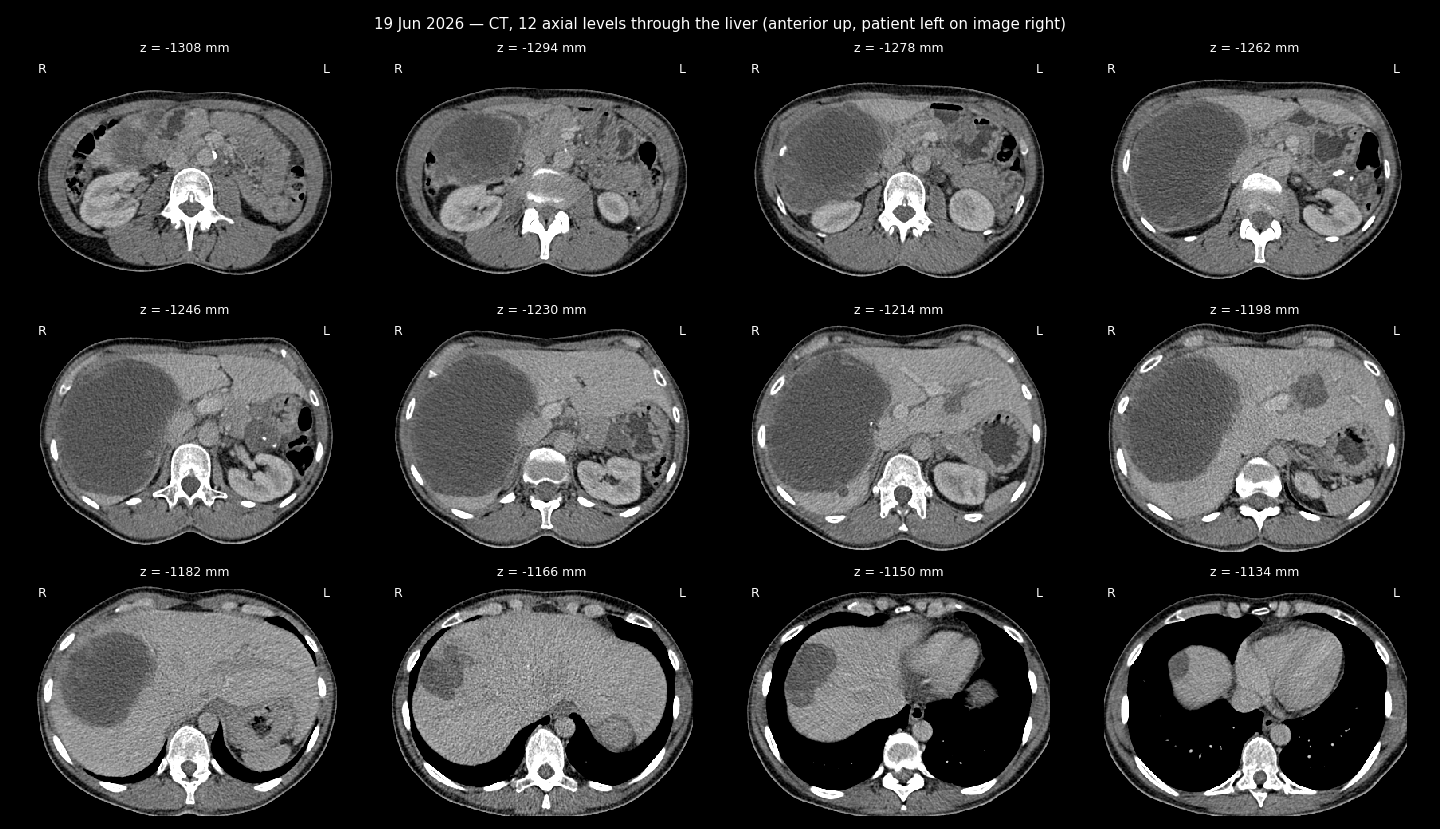
CT sheet, 12 levels through the liver
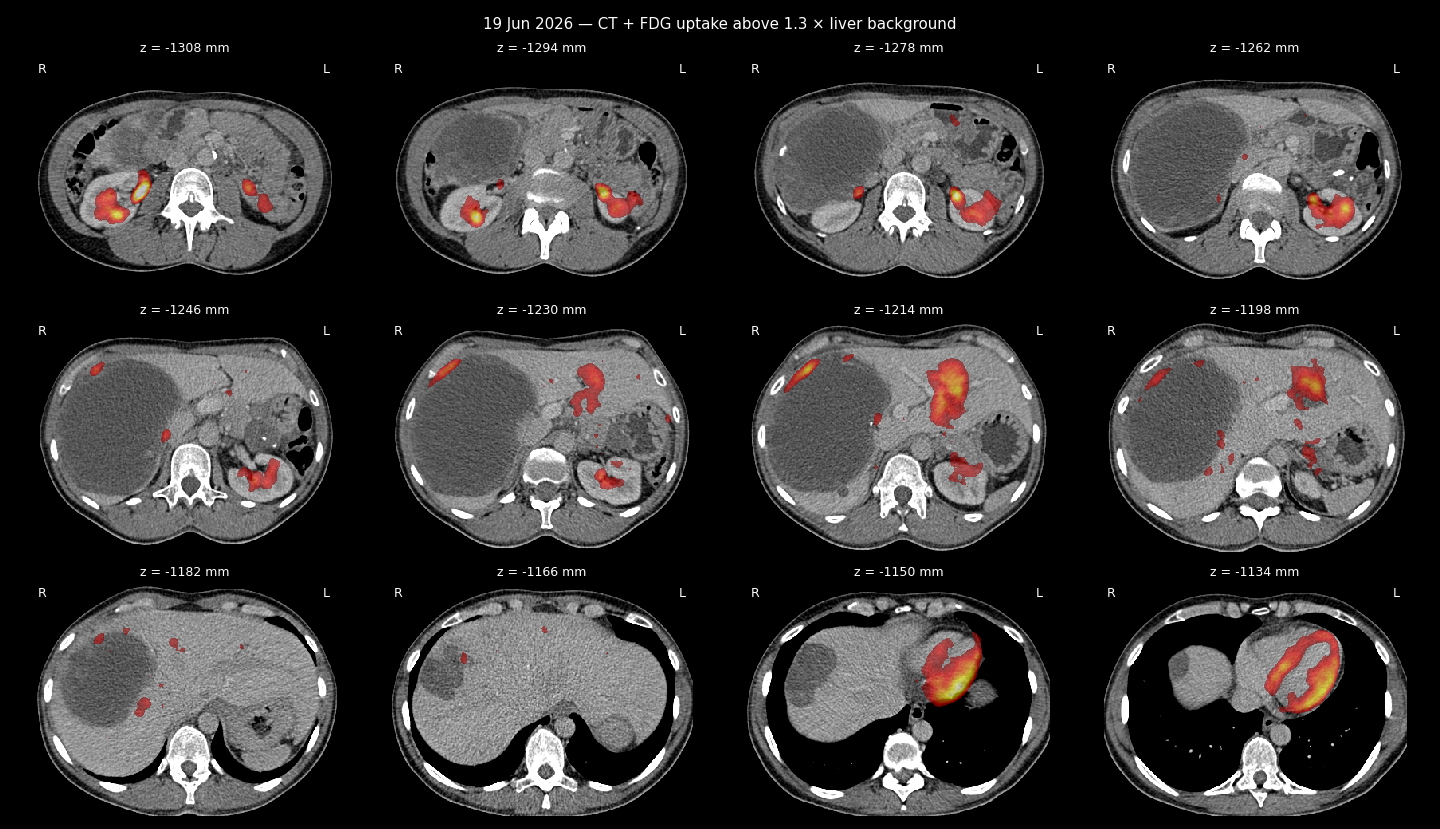
CT + FDG uptake sheet
PET-CT Whole Body with Contrast Enhancement — English Translation
Source (Ukrainian original): docs_eng/anastasia_disks/June_19_2026_PET_CT/Жерліцина_Анастасія_Станіславівна_f4b4883e_5c64_4d03_b1d1_b1cf4abb2334.pdf
Facility: All-Ukrainian Radiosurgery Center (Feofaniya), PET-CT Diagnostics Department
Patient: Zherlitsyna Anastasiia Stanislavivna, b. 1976
Study: Whole-body PET-CT with contrast enhancement
Date: 2026-06-19 / #53478/25
F18-FDG injected IV: 232.7 MBq; PET effective dose 4.4 mSv, CT 18.7 mSv
Clinical data
1. Gastrointestinal stromal tumor (GIST) of the stomach, GIST metastasis to the liver (CSD). Status post EUS-FNA biopsy of the stomach, core biopsy of the liver. 08.2023 - status after 4 months of Imatinib treatment. 03.2025 - status after 2 years of Imatinib treatment with positive clinical picture. Daily Imatinib (Glivec) continued after the last PET-CT.
2. Leiomyosarcoma of the uterine body T2NxM1, secondary involvement of liver and peritoneum. Status after 4 courses of chemotherapy per Gemcitabine+Docetaxel+Avastin protocol with disease progression in the liver (10.2022). C-KIT and MET mutations detected. Status after Pazopanib treatment (05-12.01.2023) with development of posterior reversible encephalopathy syndrome and positive CT dynamics.
01.2023 repeat liver biopsy: histology - metastasis of uterine leiomyosarcoma. Status after 3 cycles of chemotherapy per Trabectedin protocol with negative dynamics (PET-CT from 11.04.2023).
Reference PET-CT for comparison: 13.05.2025
Reference values: mediastinal blood pool SUVmax 1.41; liver parenchyma pool SUVmax 1.99.
Findings
Oncological disease control:
Stomach: not distended; along the lesser curvature a hypodense ametabolic mass persists, stable in size at up to 4.3×3.3×4.4 cm (ax+sag), with increasing calcifications in its structure.
Distant metastatic spread:
Liver: compensatorily enlarged, right lobe craniocaudal size up to 18.7 cm (13.05.2025 - up to 17.9 cm). Parenchyma contains ametabolic, hypodense masses, stable in size:
- Sg5/6/7/8: massive mass up to 14.4×10.4×14.1 cm (ax+cor)
- Sg7: satellite lesion up to 1.0×1.0 cm
- Sg7/8: subcapsular, up to 6.3×4.3 cm
- Sg5/8 border: small lesion up to 0.6×0.6 cm
- Sg4a/7 border: up to 0.6×0.6 cm
- Sg2/3 border: mass along the left portal vein, 4.5×3.6 cm
- Sg1: up to 1.3×1.3 cm
- Sg4b: small lesion up to 0.4×0.4 cm
Uterus: along the anterior wall, a heterogeneous round mass with calcifications, conditionally stable in size at up to 6.8×5.5×4.8 cm (ax+sag) - per history, likely treated leiomyosarcoma. Along the upper margin, an ametabolic mass of stable size up to 4.6×3.7 cm - pelvic MRI with contrast recommended for correct assessment. Ovaries not clearly differentiated - correlate with ultrasound data.
Bones unremarkable, no suspicious bone marrow FDG uptake.
Neck: physiological symmetric FDG metabolism in the palatine tonsils. No signs of metabolically active cervical lymphadenopathy. Thyroid not enlarged, normal structure, no increased FDG uptake. A subcutaneous port system is placed at the right subclavian region; catheter passes into the superior vena cava, distal tip at the lower third of the SVC.
Chest: mammary glands without pathological FDG uptake. No signs of metabolically active axillary, mediastinal or hilar lymphadenopathy. Trachea and main bronchi patent. Lung parenchyma normal aeration, no infiltrative or mass changes. Single small bullae on the right in S1, subpleural, up to 0.8 cm. No pleural effusion.
Abdomen/retroperitoneum: gallbladder not enlarged, compressed by adjacent liver parenchyma, contents of homogeneous density, no radiopaque calculi. Pancreas normotrophic, contour clear and scalloped. Adrenal glands not enlarged, normal shape. Kidneys typically positioned, parenchyma normal thickness. Collecting systems and ureters normal configuration. No metabolically active intra-abdominal or retroperitoneal lymphadenopathy.
Pelvis: bladder moderately filled, walls not thickened, no additional masses. Small amount of fluid in the Pouch of Douglas. No metabolically active pelvic or inguinal lymphadenopathy.
Conclusion
Compared with the PET-CT from 13.05.2025 (note: report references this as 13.05.2026, likely a typographical date error for 13.05.2025), the picture of the known gastrointestinal stromal tumor (GIST) of the stomach and metastatic liver involvement shows no reliable negative dynamics.
Given the histologically confirmed GIST, which may show absent or variable FDG avidity, the absence of pronounced metabolic activity does not exclude residual tumor tissue. For correct assessment of disease course and further monitoring, correlation with contrast-enhanced CT or MRI is recommended.
The calcified uterine body mass is stable over time; the ametabolic mass along the upper margin of the uterine body - pelvic MRI with contrast is advisable for correct assessment.
Radiologist: Zakrzhevska K.P.
Head of the All-Ukrainian Radiosurgery Center, radiologist: Kmetiuk Ya.V.
[Translation note: this is a plain-language English translation of the Ukrainian original for reference purposes; it is not a certified medical translation.]