Ultrasound of the abdominal organs and kidneys
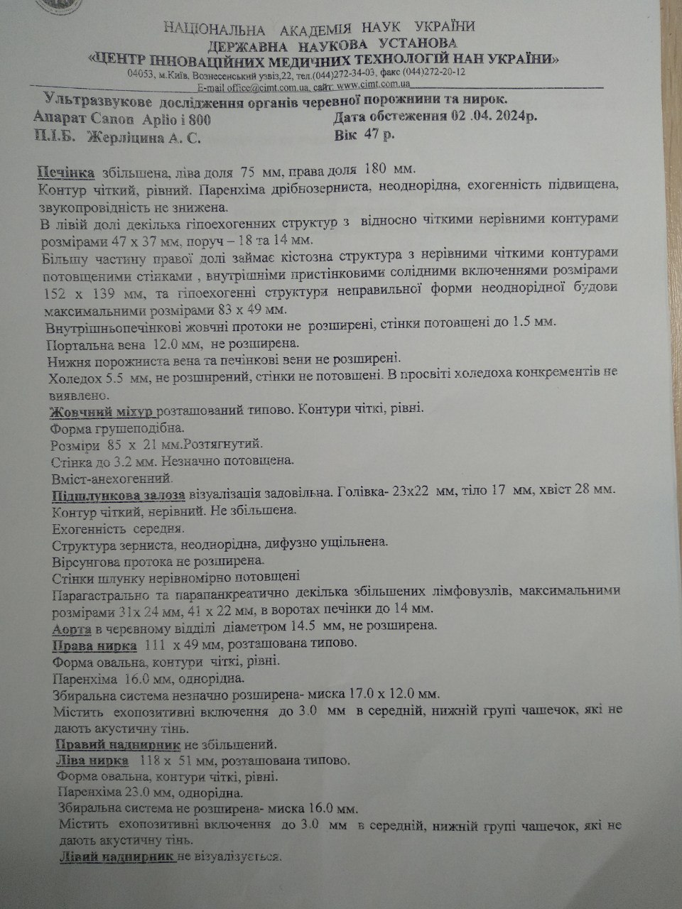
Ultrasound examination of abdominal organs and kidneys.
The liver is enlarged, the left lobe is 75 mm, the right lobe is 180 mm.
The contour is clear, even. Parenchyma is fine-grained, heterogeneous, echogenicity is increased, sound conductivity is not reduced.
In the left lobe, there are several hypoechoic structures with relatively clear uneven contours measuring 47 x 37 mm, nearby - 18 and 14 mm.
Most of the right lobe is occupied by a cystic structure with uneven clear contours and thickened walls, internal parietal solid inclusions measuring 152 x 139 mm, and hypoechoic structures of an irregular shape of heterogeneous structure with a maximum size of 83 x 49 mm.
The intrahepatic bile ducts are not dilated, the walls are thickened to 1.5 mm.
Portal vein 12.0 mm, not dilated.
The inferior vena cava and hepatic veins are not dilated.
Choledochus 5.5 mm, not dilated, walls not thickened. No concretions were found in the lumen of the choledochal.
The gallbladder is located typically. Contours are clear, even.
The shape is pear-shaped.
Dimensions 85 x 21 mm. Stretched
Wall up to 3.2 mm. Slightly thickened.
The content is anechoic.
The pancreas visualization is satisfactory. Head - 23x22 mm, body - 17 mm, tail - 28 mm.
The contour is clear, uneven. Not increased
Echogenicity is average.
The structure is granular, heterogeneous, diffusely compacted.
Virsung's duct is not dilated.
The walls of the stomach are unevenly thickened.
Paragastric and peripancreatic, several enlarged lymph nodes, the maximum size of 31 x 24 mm, 41 x 22 mm, in the gate of the liver up to 14 mm.
The aorta in the abdominal region, with a diameter of 14.5 mm, is not dilated.
The right kidney is 111 x 49 mm, located typically.
The shape is oval, the contours are clear and even.
Parenchyma 16.0 mm, homogeneous.
The collecting duct system is slightly expanded - the renal pelvis is 17.0 x 12.0 mm.
Contains echo-positive inclusions up to 3.0 mm in the middle, lower group of calyces, which do not give an acoustic shadow.
The right adrenal gland is not enlarged.
The left kidney is 118 x 51 mm, located typically.
The shape is oval, the contours are clear and even.
Parenchyma 23.0 mm, homogeneous.
The collecting duct system is not expanded - the renal pelvis is 16.0 mm.
Contains echo-positive inclusions up to 3.0 mm in the middle, lower group of cups, which do not create an acoustic shadow.
The left adrenal gland is not visualized.
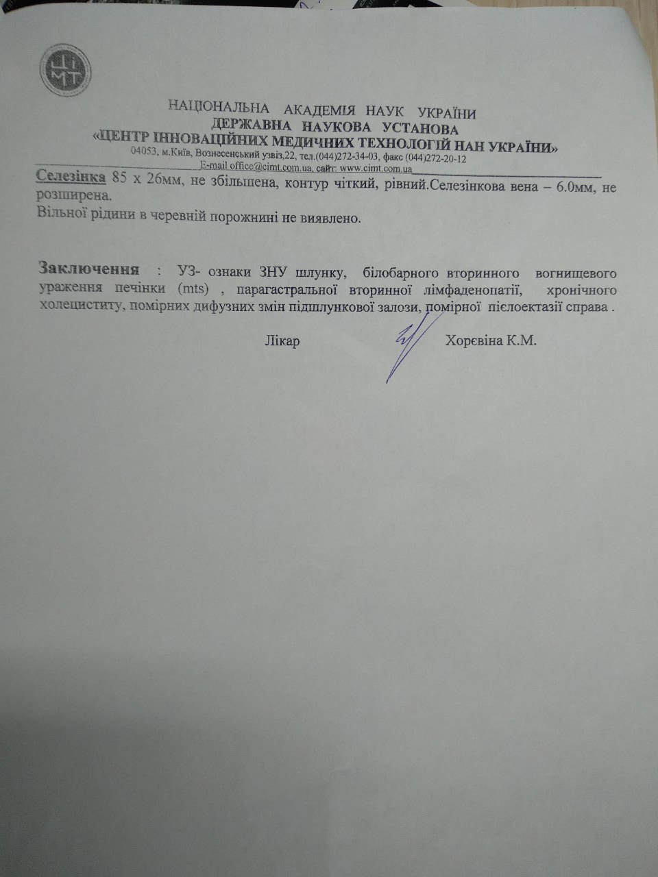
The spleen is 85 x 26 mm, not enlarged, the contour is clear, even. Splenic vein - 6.0 mm, not dilated.
No free fluid was detected in the abdominal cavity.
Conclusion: ultrasound signs of malignant neoplasms of the stomach, bilobar secondary focal liver damage (mts), paragastric secondary lymphadenopathy, chronic cholecystitis, moderate diffuse changes in the pancreas, moderate pyelectasis on the right.
| Doctor |  |
Khorevina K.M. |
|---|