MRI and MR angiography of the brain (3 T)
LLC CAPITAL
License of the Ministry
of Health of Ukraine
AG No. 599781
dated January 17, 2012
MEDICAL DOCUMENTATION
Institution code for the All-Ukrainian Classifier of Enterprises and Organizations: 32381903
Universal clinic "Oberig"
3 Zoolohichna Street, Kyiv, 03057, Ukraine
(044) 521-30-03
emergency aid 3003
5213003@oberig.ua
MRI of the brain and intra-extracranial arteries with contrast
Patient: Zherlitsyna Anastasia Stanislavivna, 46 years old
Stationary card No. 00054
Date: 20.01.2023
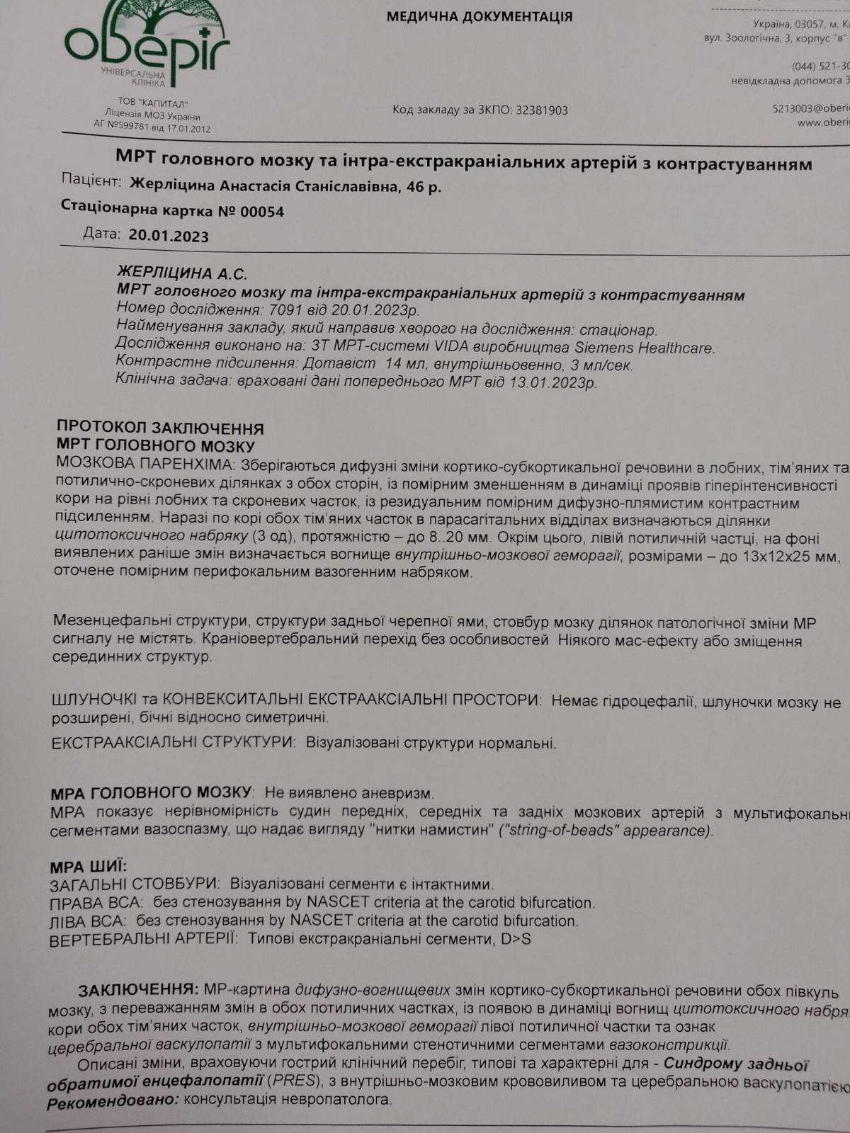
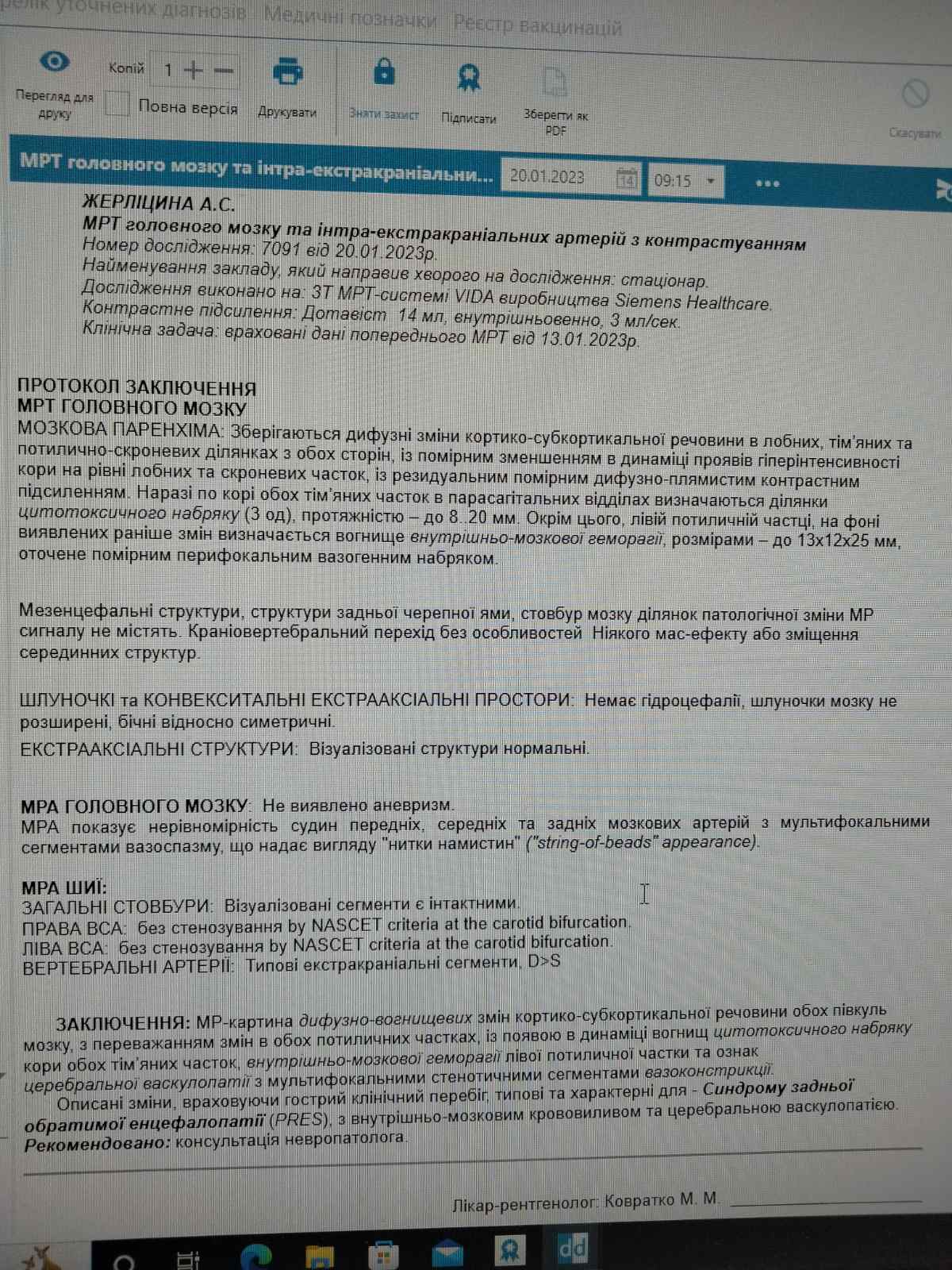
ZHERLITSYNA A.S.
MRI of the brain and intra-extracranial arteries with contrast
Research number: 7091 dated January 20, 2023.
Name of the institution that referred the patient for research: hospital.
The study was performed on: 3T VIDA MRI systems manufactured by Siemens Healthcare.
Contrast enhancement: Dotavist 14 ml, intravenous, 3 ml/sec.
Clinical task: taken into account the data of previous MRI dated January 13, 2023
PROTOCOL OF THE CONCLUSION
MRI OF THE BRAIN
BRAIN PARENCHYMA: Diffuse changes in the cortico-subcortical substance are preserved in the frontal, parietal and occipitotemporal areas on both sides, with a moderate decrease in cortical hyperintensity at the level of the frontal and temporal lobes in the dynamics, with residual moderate diffuse-spot contrast enhancement. Currently, in the cortex of both parietal lobes in the parasagittal sections, areas of cytotoxic edema (3 units) are determined, with a length of up to 8.20 mm. In addition, in the left occipital lobe, against the background of the previously detected changes, a focus of intracerebral hemorrhage, measuring up to 13×12×25 mm, surrounded by moderate perifocal vasogenic edema is determined.
Mesencephalic structures, structures of the posterior cranial fossa, and the brain stem do not contain areas of pathological changes in the MR signal. Craniovertebral transition without features. No mass effect or displacement of midline structures.
VENTRICLES and CONVEXITY EXTRA-AXIAL SPACES: No hydrocephalus, cerebral ventricles not dilated, lateral relatively symmetric.
EXTRA-AXIAL STRUCTURES: Visualized structures are normal.
MRA OF THE BRAIN: No aneurysm was found.
MRA shows irregularity of the vessels, middle and posterior cerebral arteries with multifocal segments of vasospasm, giving a "string-of-beads" appearance.
MRA OF THE NECK:
COMMON TRUNKS: Visualized segments are intact.
RIGHT INTERNAL CAROTID ARTERY: without stenosis by NASCET criteria at the carotid bifurcation.
LEFT INTERNAL CAROTID ARTERY: without stenosis by NASCET criteria at the carotid bifurcation.
VERTEBRAL ARTERIES: Typical extracranial segments, D>S
CONCLUSION: MR image of diffuse-focal changes in the cortico-subcortical substance of both hemispheres of the brain, with a predominance of changes in both occipital lobes, with the appearance in dynamics of foci of cytotoxic edema of the cortex of both parietal lobes, intracerebral hemorrhage of the left occipital lobe and signs of cerebral vasculopathy with multifocal stenotic segments vasoconstriction.
The described changes, taking into account the acute clinical course, are typical and characteristic of - Posterior Reversible Encephalopathy Syndrome (PRES), with intracerebral hemorrhage and cerebral vasculopathy.
Recommended: consultation of a neurologist.
Radiologist: Kovratko M. M_________________